


方案详情
文
过氧化氢是一种主要的氧化还原信号分子,是细胞功能和通讯的新范式的基础。H2O2作为细胞间信号分子和神经调节剂在大脑中的作用越来越明显,证据表明这种生物氧化剂可以调节神经元的极性、连接性、突触传递和神经元网络的调节。这一概念得到了其在细胞外空间(从生产源到目标)扩散的能力的支持。因此,了解活体大脑中细胞外H2O2浓度动态以及影响其扩散模式和半衰期的因素至关重要。为了解决这个问题,本文使用了一种新的微传感器来测量脑细胞外基质中H2O2的浓度动态,无论是在使用啮齿动物脑切片的离体模型中还是在体内。本文研究人员发现外源施加的H2O2从细胞外空间中去除,体内平均半衰期为t1/2=2.2 s,并确定H2O2的体内有效扩散系数为D*=2.5×10⁻⁵ cm2 s−1。这使其在半衰期内在细胞外空间扩散超过100μm。考虑到这一点,可以暂时将H2O2放在体积神经递质的类别中,连接大脑组织复杂网络中的所有细胞类型,无论它们是否物理连接。大脑中H2O2扩散和半衰期的这些定量细节使我们能够解释氧化还原信号的生理学,并为解决与疾病过程相关的氧化还原稳态失调奠定基础。
方案详情

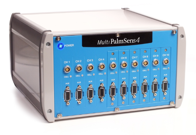
过氧化氢是一种主要的氧化还原信号分子,是细胞功能和通讯的新范式的基础。H2O2作为细胞间信号分子和神经调节剂在大脑中的作用越来越明显,证据表明这种生物氧化剂可以调节神经元的极性、连接性、突触传递和神经元网络的调节。这一概念得到了其在细胞外空间(从生产源到目标)扩散的能力的支持。因此,了解活体大脑中细胞外H2O2浓度动态以及影响其扩散模式和半衰期的因素至关重要。为了解决这个问题,本文使用了一种新的微传感器来测量脑细胞外基质中H2O2的浓度动态,无论是在使用啮齿动物脑切片的离体模型中还是在体内。本文研究人员发现外源施加的H2O2从细胞外空间中去除,体内平均半衰期为t1/2=2.2 s,并确定H2O2的体内有效扩散系数为D*=2.5×10⁻⁵ cm2 s−1。这使其在半衰期内在细胞外空间扩散超过100μm。考虑到这一点,可以暂时将H2O2放在体积神经递质的类别中,连接大脑组织复杂网络中的所有细胞类型,无论它们是否物理连接。大脑中H2O2扩散和半衰期的这些定量细节使我们能够解释氧化还原信号的生理学,并为解决与疾病过程相关的氧化还原稳态失调奠定基础。 原文链接:https://doi.org/10.1016/j.redox.2022.102250本文中进行电化学测试的仪器为荷兰PalmSens,型号:MultiPalmSens4多通道电化学分析仪,测试方法为FCV(快速循环伏安扫描法)。Redox Biology 50 (2022) 102250ELSEVIERContents lists available at ScienceDirect A. Ledo et al.Redox Biology 50 (2022) 102250 Redox Biology journal homepage: www.elsevier.com/locate/redox In vivo hydrogen peroxide diffusivity in brain tissue supports volumesignaling activity A. Ledo a b,", E. Fernandes , A. Salvador D,,cc,,dd,, J. Laranjinha a,b,R.M. Barbosaa,b Faculty of Pharmacy, University of Coimbra, Azinhaga de Santa Comba, 3000-548, Coimbra, Portugal DCenter for Neuroscience and Cell Biology, University of Coimbra, Rua Larga, 3004-504, Coimbra, Portugal C Institute for Interdisciplinary Research, University of Coimbra, Casa Costa Alemāo, 3030-789, Coimbra, Portugal “CQC, Department of Chemistry, University of Coimbra, Rua Larga, 3000-535, Coimbra, Portugal ARTICLEINFO ABSTRACT Keywords:Hydrogen peroxideVolume signalingDiffusionBrain Hydrogen peroxide is a major redox signaling molecule underlying a novel paradigm of cell function andcommunication. A role for H202 as an intercellular signaling molecule and neuromodulator in the brain hasbecome increasingly apparent, with evidence showing this biological oxidant to regulate neuronal polarity,connectivity, synaptic transmission and tuning of neuronal networks. This notion is supported by its ability todiffuse in the extracellular space, from source of production to target. It is, thus, crucial to understand extra-cellular H202 concentration dynamics in the living brain and the factors which shape its diffusion pattern andhalf-life. To address this issue, we have used a novel microsensor to measure H2O2 concentration dynamics in thebrain extracellular matrix both in an ex vivo model using rodent brain slices and in vivo. We found that exoge-nously applied H202 is removed from the extracellular space with an average half-life of t1/2=2.2 s in vivo. Wedetermined the in vivo effective diffusion coefficient of H202 to be D*=2.5×10⁻⁵ cm’s-. This allows it todiffuse over 100 um in the extracellular space within its half-life. Considering this, we can tentatively place H202within the class of volume neurotransmitters, connecting all cell types within the complex network of braintissue, regardless of whether they are physically connected. These quantitative details of H202 diffusion and half-life in the brain allow us to interpret the physiology of the redox signal and lay the pavement to then addressdysregulation in redox homeostasis associated with disease processes. 1. Introduction Hydrogen peroxide - H202 - is a major biological oxidant whichplays a pleiotropic role in redox regulation of cell functions. Its intra-cellular concentration is maintained in the steady-state low nanomolarrange through regulation of production and efficient redundant removalsystems [1]. H202 is a strong 2-electron oxidant, but its high activationenergy renders it relatively stable and limits its reaction with mostbiological molecules. In the brain, this oxidant has been shown toregulate neuronal polarity, connectivity, synaptic transmission in addi-tion to tuning of neuronal networks (reviewed in Refs. [2,3]), and redoximbalance or oxidative stress are strongly associated with neuro-degeneration and can undermine deleterious neuroinflammatory re-sponses [4]. In the striatum H2O2 is proposed to be released frommedium spiny neurons in response to glutamate depolarization, result-ing in the down-regulation of dopamine release from midbrain afferents (reviewed in Ref.[5]). The intracellular concentration of H202 is maintained in low nMlevels due to the activity of molecular sinks [6,7], while extracellularconcentrations may be higher. Some reports point towards a bloodplasma level as high as 1-5 uM [8], however careful experiments andcritical analysis suggest that blood plasma levels of H202 may be in thelow nM range, with the exception of situations such as inflammation orinfection [9-11]. Also, the concentration of H2O2 varies in mammaliancells within different compartments: lowest [H2O2] is found in thecytosol (80 pM-2.2 nM), while inside the endoplasmic reticulum, 700nM concentration has been reported [6,12,13]. Steep concentrationgradients are expected to be found in the cytosol centered at H2O2 entryor production sites [13,14]. Although H202 can cross biological mem-branes through passive diffusion [15,16], sharp gradients across bio-membranes were identified in the seminal work by Antunes and Cadenas[17]. Higher transport rates are afforded by several aquaporins, coined * Corresponding author. Faculty of Pharmacy, University of Coimbra, Azinhaga de Santa Comba, 3000-548, Coimbra, Portugal. 2213-2317/C 2022 The Authors. Published byyElsevierB.V. This is an open access article under the CCFBY-NC-ND license(http://creativecommons.org/licenses/by-ne-nd/4.0/). “peroxiporins”, shown to facilitate H2O2 diffusion across biomembranes[18]. The occurrence of H202 transport systems across the cell mem-branes advances the notion that the extracellular concentration of H202impacts dynamically on intracellular signaling pathways in neighboringcells. Enzymes responsible for removal of H202 include peroxiredoxins(Prx) and glutathione peroxidases (GPx), both of which display highsecond-order rate constants in the range of 10-10° M-1s-l relative toH2O2 [19]. Peroxiredoxins can transmit the oxidizing equivalents toother target proteins, acting as redox relays in thiol-dependent H202signaling [20]. Catalase (CAT) is a heme protein that, in its main cata-lytic cycle, catalyzes the 2-step dismutation of two H202 molecules towater and O2 with no net consumption of cellular reducing equivalents[21,22]. Finally,peroxidase enzymes use H202 as a substrate to generateoxidants, as is the case of myeloperoxidase found in neutrophils thatproduces hypochlorous acid as part of the pathogen defense mechanism[23]. Inherent to the notion of H202 as an intercellular signaling moleculeis its diffusion in the extracellular space. To understand the distributionand range of influence of a particular diffusible compound in brain tissueone must know its effective diffusion coefficient (D*) and the relativeimportance of diffusion vs clearance processes that participate in theremoval and or degradation/decomposition from the extracellular space(ECS). Diffusion in the ECS can be modified by removal across the bloodbrain barrier, uptake into cells or binding to receptors (conceptsreviewed in Ref. [24]). Here we have investigated H202 concentrationdynamics in the brain ECS in the striatum both in an acute slice prep-aration and in vivo. To this purpose we have used novel rutheniumpurple modified carbon fiber microelectrodes (RP-CFM) which we havepreviously designed and characterized for their electrocatalyticalproperties towards the selective detection of H202 concentration withhigh spatial and temporal resolution [25]. We determined the half-life ofH202 in brain tissue and used mathematical modelling to determine D*in vivo, thus inferring its diffusional spread in the brain. 2. Materials and methods Reagents and Solutions: Reagents were analytical grade and ob-tained from Sigma-Aldrich. Argon was provided by Air Liquide, Portugaland Carbox (95% 02/5%CO2) was obtain from Linde Sogas, Portugal.All solutions were prepared in ultra-pure deionized water (≥18 MQ cm)from a Milli-Q system (Millipore Company, Bedford, MA, USA). Micro-electrode in vitro evaluations were performed in phosphate bufferedsaline (PBS) with the following composition (in mM): 140 NaCl, 2.7KCl,8.1 Na2HPO4, 1.8 KH2PO4, pH 7.4. Media for hippocampal slice exper-iments was artificial cerebrospinal fluid (aCSF) composed of (in mM):124 NaCl, 2 KCl, 25 NaHCO3, 1.25 KH2PO4, 1.5 CaCl2 and 10 p-glucose.For dissection and recovery, a modified aCSF was used to increaseviability. Composition of this aCSF was (in mM): 124 NaCl, 2 KCl, 25NaHCO3, 1.25 KH2PO4, 0.5 CaCl2, 10 MgSO4, 0.2 AA, 1 GSH and 10 D-glucose. In all cases, aCSF was continuously bubbled with humidifiedCarbox for pH buffering (pH 7.4) and oxygenation. Animals: All the procedures in this study were performed in accor-dance with the European Union Council Directive for the Care and Use ofLaboratory animals, 2010/63/EU, and were approved by the local ethicscommittee (ORBEA) and the Portuguese Directorate-General for Foodand Veterinary. A total of 21 male Wistar rats aged 8-12 weeks andweighing 300-390 g (Charles-River Laboratories, Barcelona, Spain)were used in these experiments: 6 for slices and 15 for in vivo. While inthe animal facility, animal husbandry conditions were as follows:housed in pairs in filter-topped type III Makrolon cages in the local vi-varium with controlled environmental conditions, namely a tempera-ture of 22-24°C, relative humidity of 45-65%, air exchange rate of 15times per hour, 12 h light/dark cycle, and with standard rat chow diet(4RF21-GLP Mucedola, SRL, Settimo Milanese, Italy) and chlorinatedwater available ad libitum. Carbon Fiber Microelectrode Fabrication, Modification andEvaluation: Carbon fiber microelectrodes (CFM) were fabricated aspreviously described [26]. Briefly, a single carbon fiber (30 um o.d.;Textron Lowell, MA, USA) was inserted into a borosilicate glass capillary(1.16 mm i.d. and 2.0 mm o.d.; Harvard Apparatus, Holliston, MA,USA)and cleaned with acetone. Each capillary was pulled using a verticalpuller (Harvard Apparatus, UK) and the protruding carbon fiber was cutto a tip length of approx. 150 um. The electrical contact between thecarbon fiber and the copper wire was provided by conductive silverpaint (RS, Northants, UK). The microelectrodes were tested for generalrecording properties in 0.05 M PBS Lite (in mM: 10 Na2HPO4, 40NaH2PO4, and 100 NaCl, pH 7.4) by fast cyclic voltammetry at a scanrate of 200 V s-l, between -0.4 and + 1.6 V vs Ag/AgCl for 30 cycles. Ruthenium purple (RP) modification of CFM was achieved asdescribed in Ref. [25]. Briefly, a solution of 1 mM K4 [Ru"(CN)6] pre-pared in 35 mM KCl (N2-purged) was mixed, under vigorous stirring,with a solution of 1 mM FeClg in 35 mM KCl (N2-purged). The pH of theresulting colloidal suspension was adjusted to 2.0 using 1 M HCl. Thissolution was placed in an ultrasound bath and RuCl3 was added from a 1mM solution to give a final concentration of 20 uM. This solution wasused within 4 h of preparation. Electrodeposition was carried out bypotential cycling between -0.2 and + 1.0 V vs Ag/AgCl at a scan rate of50 mV s-. The cycles were repeated until the redox peaks no longerincreased in height. The modified CFM (CFM-RP) was rinsed withdistilled water and placed in a solution of 1 mM RuCl3 in 35 mM KCl.Between 2 and 4 further cycles were performed under the same condi-tions to ensure the stability of the deposited film. For coating withNafion@, the tip of the CFM-RP was dipped into a 5% solution ofNafionQ in aliphatic alcohols for 5 s and then dried at 100 °C for 15 min.These sensors were designated CFM-RP-Nafion@. Preparation of Rat Striatum Slices: Following decapitation underdeep anesthesia (isoflurane), the brain of the animal was rapidlyremoved and placed in ice-cold, Carbox bubbled modified aCSF. Thecerebellum was removed, the two hemispheres were separated andmounted in the pre-chilled stage of a vibratome (Vibroslice, WorldPrecision Instruments) and submerged in the chamber filled with icecold modified aCSF continuously bubbled with Carbox. Brain slicescontaining the striatum were obtained with a thickness of 400 um andtransferred to a pre-incubation chamber (BSC-PC; Harvard Apparatus)filled with modified aCSF. Slices were allowed to recover under theseconditions for at least 1 h prior to recording. Recording H202 in Rat Striatal Slices: An individual slice wasplaced in a recording chamber (BSC-BU with BSC-ZT top, HarvardApparatus) and perfused with aCSF continuously bubbled with humid-ified Carbox and at a flow rate of 1.5-2 mL min-. The temperature ofthe chamber was maintained at 32 °C (temperature controller model TC-202A, Harvard Apparatus). A recording array comprised of a CFM-RP-Nafion@ and a CFM mounted at a tip-to-tip distance of 100 um waslowered into the tissue with the aid of a micromanipulator so as toguarantee that the totality of the active surface was in the tissue core.Recording of the reduction current was initiated and once a stablebaseline was obtained, an H202 solution (1 mM in aCSF) was pressureejected (10 Psi, 1s) via a Picospritzer II (General Valve, Fairfield, NJ)through a pulled micropipette, placed between the two workingelectrodes. 2.1. In vivo recording of hydrogen peroxide in the brain of anesthetizedrats The experimental setup used for amperometric monitoring of H202in vivo in anesthetized rats was similar to that used in previous studies[27]. Briefly, an animal was anesthetized with urethane 1.25 gkg(i.p.)and placed in a stereotaxic apparatus. Body temperature was maintainedat 37 °C with a heated pad coupled to a Gaymar Heating Pump (Brain-tree Scientific, Inc., USA). The skull was exposed by a midline scalpincision and retraction of the skin and temporal muscle. Bleeding was controlled using a Bovie@ cautery. A craniotomy was made over thecerebral cortex with an area of roughly 7 mm² (AP: +3.0 to +0.5 mm;ML: ±1 to ±4 mm; relative to bregma [28]) with removal of the overlying meninges. An additional small burr-hole was drilled in a siteremote from the recording area for the insertion of a miniature Ag/AgClreference electrode in the subdural space. The cortical surface wasmaintained wet with saline soaked cotton balls. An array comprising aCFM-RP-NafionQ and a pulled micropipette (average distance of 75 ±10 um) was lowered into the dorsal medial striatum (AP: +1.5-2.0 mm;ML: ±1.5-±2.5 mm and DV: -4.0 to -6.0 mm relative to bregma).After insertion of the array into the brain, the baseline was allowed tostabilize for at least 20 min. The mean display frequency was set at 4-20Hz. An H202 solution (1 mM in aCSF) was pressure ejected via aPicospritzer II (General Valve, Fairfield, NJ) through a pulled micropi-pette, with a volume dispensed of 3-125 nL. In experiments in whichcardiac arrest was performed during recording session, the rat received alethal dose of euthanasia solution (0.5 mL of sodium pentobarbital 200g mL-,i.p.). Electrochemical Instrumentation: Electrochemical proceduresincluding RP electrodeposition and CFM evaluation by FCV were per-formed using aMulti PalmSens4Potentiostat (PalmSens, TheNetherlands) controlled by MultiTrace v.4.2 software (PalmSens, TheNetherlands). A 3-electrode electrochemical cell was used, comprised ofthe working electrode, an Ag/AgCl in 3 M NaCl reference electrode (RE-5B, BAS Inc., IN, USA), and a Pt wire as auxiliary electrode. Ampero-metric recording in slices and calibration in the slice recording chamberwere performed using a FAST16mkII potentiostat (Quanteon, KY, USA)in a 2-electrode electrochemical cell configuration comprised of theworking electrode and an Ag/AgCl pellet reference electrode. Ampero-metric recording in vivo in the anesthetized rodent brain was performedusing a FAST16mkIII potentiostat (Quanteon, KY, USA) in a 2-electrodeelectrochemical cell comprised of a working electrode and an Ag/AgClminiature pseudo reference electrode. The latter was produced byanodization of the exposed tip of the Teflon-coated Ag wire in 1 M HClsaturated with NaCl, which, when in contact with cerebrospinal fluid inthe brain containing chloride ions, develops an Ag/AgCl half-cell. BothFAST16 potentiostats were controlled by FAST2014 software (Quan-teon, KY, USA). Working potential for monitoring H202 was set at -0.2V vs Ag/AgCl [25]. Data Analysis. Data analysis was performed using MultiTrace v.4.2,FAST Analysis version 6.0, OriginPro 2016, and GraphPad 5.0. Valuesare given as the mean ± SEM unless otherwise stated. Normality of datawas confirmed using the D'Agostino and Pearson omnibus normality test(α=0.05). Calculated parameters were statistically evaluated by usingOne-way ANOVA followed by Dunnett post-hoc test. The number ofrepetitions is indicated in each individual determination. The sensitivityof CFM-RP-Nafion@ toward H202 reduction was determined by linearregression analysis in the range of 0-50 pM. The diffusion of H202 from aspherical source was modeled using OriginPro 2016, based on equation(1), which describes diffusion from a spherical source [30]. 3. Results 3.1. Uptake and removal ofextracellular Hydrogen Peroxide in rat brainstriatum slices We evaluated H2O2 concentration dynamics in rat striatum slicesusing previously described CFM-RP-Nafion@ sensors [25] in order toinvestigate how brain tissue handles an increase in extracellular H202-To this purpose, we constructed an array composed of a H202 sensor anda null sensor with a tip-to-tip distance of ca. 100 um. This array waslowered into the tissue core, in the dorsal-medial region of the striatalslice. A micropipette with a tip diameter of 10-20 um and filled with a 1mM H2O2 solution was placed between the 2 sensors and a volume ofsolution (25-50 nL) was pressure ejected for a period of 1 s, as depictedin the inset of Fig. 1B. As can be observed in Fig. 1A, repeated H202 by exogenous local application of a 1 mM H2O2 solution. Blue trace representsthe amperometric current measured at the CFM-RP-NafionQ H2O2 sensor whilethe grey trace represents the current recorded at a null sensor. Repeatedapplication of H202 through a micropipette place between the 2 sensors pro-duced transient and reproducible increase in H202 in the brain tissue. B -Average of 44 individual normalized signals (blue) and first order exponentialdecay function (red). Inset is a schematic representation of the experimentaldesign used, including the microelectrodes and ejection pipette between thetwo, where d is the distance between the micropipette tip and each of thesensors, v is the volume of the H2O2 solution ejected from the micropipette andr is the calculated radius of the bolus of H202 ejected. (For interpretation of thereferences to colour in this figure legend, the reader is referred to the Webversion of this article.) ejections (3 min inter-pulse interval) resulted in transient increases inH2O2 in the tissue recorded at the CFM-RP-NafionQ sensor, with nochange in the current recorded at the null sensor. The decay phase of the transient H202 signal was suitably fitted to a1st order exponential decay function, shown as the superimposed redline over the experimental data in blue circles in Fig. 1B. From thisfitting we determined the time constant of the signal decay,k=0.32±0.02s-, as well as the decay half-time,t1/2=2.5±0.1 s (N =44 signals,obtained from 6 slices). In order to discriminate the role of brain tissue tortuosity vs intra-cellular processes governing H2O2 uptake and breakdown, we repeated the above experiment in a matrix mimicking tissue tortuosity but devoidof cellular components. We obtained 400 um slices from a 0.2% agaroseblock [29,30], which were placed in the slice recording chamber andevaluated H2O2 signal obtained as with striatum slices. As summarizedin Table 1, the recorded H202 signals showed slower decay kinetics andthe time constant of H202 decay was significantly decreased ascompared to that found in striatal slices (k=0.11±0.01 s-l) with in-crease in the signal half-time (t1/2=6.5±0.3 s, N=5, p<0.0001).Additionally, we determined the H2O2 signal profile in metabolicallycompromised striatum slices perfused in 0 mM Glucose, 10 mM Sac-charose and 10 mM CN . Under these conditions, the H202 signal wassimilar to that observed in agarose slices and the decay time constant (k= 0.12 ± 0.01 s-) and signal half-time (t1/2=5.8±0.5 s) weresignificantly changed as compared to control tissue (p <0.0001). Theseresults support a role for cellular components in shaping extracellularH2O2 concentration dynamics. 3.2. Hydrogen Peroxide removal In Vivo in the rat striatum Slice preparations preserve tissue cytoarchitecture and allow us tounderstand the role of neural cells (neurons and astrocytes) in modu-lating the concentration dynamics of diffusible species such as H202.However, they are devoid of functional vasculature. Hence, we inves-tigated H202 signal profiles in vivo in the striatum of anesthetized rats.Similar to what was previously observed in striatal slices, local pressureejection of a 1 mM solution resulted in a transient increase in H202recorded at the CFM-RP-NafionQ H202 sensor inserted in the dorsalmedial striatum in vivo in the anesthetized rat brain and repetitiveejections produced reproducible signals in vivo (Fig. S1). The decay phase of the signal was well adjusted to a 1s orderexponential decay function (Fig. 2A) and average time constant wasdetermined to be k= 0.34±0.01 s, corresponding to an average t1/2=2.2±0.1s(N= 121 signals, obtained from 12 recording sessions,Table 1), similar to that observed in slices. Furthermore, the averagepeak concentration of H202 measured at the detector was 30.5±2.5uM.We repeated this paradigm using a higher concentration of H202(10mM) and observed that the average time constant significantlydecreased to k = 0.20±0.02s-(N = 43 signals, obtained from 3recording sessions, Table 1) and the average H202 peak concentrationwas increased to 105.9±30.4 uM. This result suggests that for 10 mMH202, the Prx pool becomes partially hyperoxidized [31], which mighthinder its participation in H202 removal, resulting in slower H202clearance by the tissue. For the remainder of the experiments we used a1 mM H202 solution. Induction of cardiac arrest resulted in rapid loss of spontaneous brainwave activity accompanied by a gradual decrease of the k values Table 1 Comparison of decay time constant (k) and half-life (t1/2) of H202 signals ob-tained in response to puff ejections of a 1 mM H202 solution, unless otherwiseindicated. Data were obtained in ex vivo acute striatal slices and 0.2% agaroseslices, and in vivo in the dorsal medial striatum in anesthetized rat and in a 0.2%agarose block at 37 °C. Values represent mean ± SEM. *p < 0.0001 as comparedto control slices and #p < 0.001 as compared to in vivo control (1 mM H202). Experimental Model (N) k/s t1/2/S Ex vivo - Striatum Slices Control (44) 0.32±0.02 2.5±0.1 MC (5) 0.12±0.01* 5.8±0.5* Agarose 0.2%(5) 0.11±0.01* 6.5±0.3* In vivo - Dorsal Medial Control (121) 0.34±0.01 2.2±0.1 Striatum 10mM H202 (42) 0.23±0.02” 3.5±0.2* A.D.(19) 0.21±0.03* 4.8±0.7* Agarose 0.2% 0.04土 18.6± (26) 0.004* 1.2” MC - metabolically compromised slices, perfused with 0 mM glucose, 10 mMsaccharose and 10 mM CN. A.D. - after death evoked by lethal dose of pentobarbital. (Fig. 2B). As summarized in Table 1, the average time constant valuestabilized at k = 0.21±0.03 (N= 19), a value significantly lower whencompared to the control situation, but similar to that observed in slicesfollowing metabolic poisoning (p=0.87) and in a 0.2% agarose block (p=0.83) used as a mimetic of brain tissue tortuosity. The onset ofdecrease in k values coincided with the decrease in power observed inthe spectrogram, known to reflect brain wave activity [32-34]. 3.3. Estimation of Hydrogen Peroxide diffusion coefficient in vivo in thestriatum In order to determine de in vivo effective diffusion coefficient (D*) ofexogenously applied H202 in striatum tissue, the experimental data wereinput to Eq. (1) where C and Co represent the instantaneous and initialconcentration, r is the radius of the sphere of H2O2 ejected, d is thedistance between source (pipette) and recorder (H202 sensor), t is time,D* the effective diffusion coefficient and 人 represents the inactivationconstant. (1) The experimental design was adjusted to guarantee r < d, as illus-trated in Fig. 3B. Recordings were obtained both in vivo, in the striatumof anesthetized rats and from a 0.2% agarose gel block at 37°C. Asshown in Fig. 3A, in both cases we observed a good adjustment betweenthe experimental data (open circles) and the fit to Eq. (1) (line). We determined the D*in vivo of H202 to be (2.5±0.1)x10-5cm²s-1with an average inactivation constant of 人= 0.19±0.01s-(N=57obtained from 5 recording sessions). Interestingly, this value of D* wasfound to be significantly higher than that calculated from data obtainedin agarose gel, where we observe D*agarose =(1.7±0.1×10⁻⁵)cm²s-1and an inactivation constant of 人=0.005±0.002s-(N=38). From the Einstein-Smoluchowski Eq. (2) one can define the averagesquare of the distance travelled (x2) by an individual molecule as afunction D and t [35]. As can be appreciated in Fig. 4, H202 can beclassified in the same range as small volume neurotransmitters such asnitric oxide ("NO), displaying a relatively large displacement within itst1/2 (112 um) when compared to other neurotransmitters such asdopamine or glutamate (properties summarized in Table 2). 4. Discussion Interest in monitoring H202 in brain tissue extracellular space hasgrown over the past years as a result of increasing recognition of itsinvolvement not only in the etiology of neurodegenerative disorderssuch as Parkinson’s and Alzheimer's diseases [39], but also due to theappreciation of its putative role as a diffusible neuromodulator, analo-gous to *NO [5,40]. Recent advances in the identification of membraneH2O2 transporters, beyond the passive diffusion through membranes,support that the extracellular H2O2 dynamics might modulate intracel-lular redox signaling pathways in cells localized within the volume ofinfluence of H202. Here, we have investigated the extracellular con-centration dynamics of H202 in the striatum, a brain region where H202has been proposed to act as an intercellular messenger molecule,modulating dopamine release from midbrain afferent fibers [41,42],focusing on determining H202 half-life time in brain tissue in an ex vivomodel (striatal slices) and in vivo in the striatum of anesthetized rats. Inaddition, we have determined the effective diffusion coefficient of H202in brain tissue in vivo. We used an artificial peroxidase type electro-chemical microsensor [25] to monitor real-time H202 concentrationdynamics in the brain extracellular space with high sensitivity andselectivity towards H2O2 and high spatial and temporal resolution version of this article.) resulting from the reduced sensor size (o.d. 33 um) and the fast-samplingability of amperometry. In both experimental models, we coupled amicropipette to the microsensor to apply a controlled pressure ejectionof a nL volume of a H2O2 solution at a known distance from the detector.Through mathematical fitting of the data, to the best of our knowledge,we provide the first in vivo data regarding the effective or apparentdiffusion coefficient of H202 in the living brain. Hydrogen Peroxide is Rapidly Removed from the Extracellular Space by Metabolically Active Cells. In the first set of experiments, weinvestigated the extracellular concentration dynamics of H202 in an exvivo model of acute striatum slices. This experimental model has theadvantage of preserving the brain tissue cytoarchitectural integrity,namely connectivity and intercellular communication. Because cere-brovascular blood flow is absent in this model, it allows us to betterunderstand the role of neural cells (astrocytes and neurons) without thecontribution of blood flow. Our data revealed that a puff application of a H2O2 solution in the proximity of the detector resulted in a transientincrease in H202 concentration, revealing that extracellular H202 dif-fuses away from the point of origin and is rapidly removed. Removal ofexogenously applied H202 was found to be the result of intracellularfactors, as the decay was significantly slower in a 0.2% agarose slice.Agarose has been widely used to mimic brain tissue tortuosity and toevaluate its effect on the diffusion of chemical species [29,30]. In linewith this observation, in metabolically compromised striatal slicesperfused with CN~ and 0 mM glucose as a strategy to hinder energymetabolism, H202 decay was found to be similar to that observed inagarose. Taken together, these results support the notion that Data (In vivo) D*= 2.6x10cm²s Fig. 3. -A-Representative recordings of H202 concentration dynamics obtained in vivo in the striatum (blue-green) and in a 0.2% agarose block at 37°C (grey).Circles represent experimental data while lines correspond to the theoretical trace obtained by modelling expression (1). B-Experimental design used, comprising amicropipette filled with a 1 mM H202 solution coupled to a CFM-RP-Nafion@ sensor for H202 detection. Interval of distance (d) between source and detector as wellas for the volume and radius of applied H202 bolus are discriminated. As drawn, d=300 um and r=100 um. C-Effective diffusion coefficient values calculated bymodelling experimental data with Eq. (1). (For interpretation of the references to colour in this figure legend, the reader is referred to the Web version of this article.) extracellular H202 enters cells, namely neurons and/or astrocytes, and isremoved in metabolically competent cells, presumably through the ac-tivity of enzymatic systems such as GPx, Prx and CAT. Similarly, in vivo, we also observed that puff application of an H202solution resulted in a transient increase in extracellular H202, with adecay rate superior to that found in a 0.2% agarose block. Followingcardiac arrest, the decay rate of the signal decreased progressively, butremained above that found in agarose. We speculate that this is theresult of H202 removal by erythrocytes, namely due to the activity ofcatalase which does not consume redox equivalents when degradingH2O2 [43]. While in neurons and astrocytes, catalase is mainly found in t/s Fig. 4. Calculated root-mean-square (R.M.S.) dis-tance of random movement for H202 using Eq. (2)and considering D* values calculated in presentstudy. For comparative purposes we have alsoplotted data for NO (grey), dopamine (DA, blue),NO2 (orange) and glutamate (purple). Red circlesrepresent distance travelled by each analyte for theirrespective extracellular half-life time (t1/2). (Forinterpretation of the references to colour in thisfigure legend, the reader is referred to the Webversion of this article.) Table 2 Comparative analysis of half-life and apparent diffusion coefficient for H2O2 andother species acting as volume transmission molecules (*NO and dopamine) aswell as the fast-excitatory neurotransmitter glutamate. Nitrite has been includedto show the effect of charge on a small molecule. Analyte Experimental Model t1/2 D*(x10~ Ref (s) cm²s-1) H202 In vivo in the rat striatum 2.2 2.5 Present Study NO In vivo in the rat cortex 0.6 3.3 [36] Dopamine Midbrain slices from 8.1 0.69 [37] Nitrite Guinea pig1g n.d. 0.63 [361 Glutamate Mathematical Modelling n.d. 0.25 [381 peroxisomes and thus less accessible to exogenously derived H202,erythrocytes do not have membrane organelles and CAT is found in thecytosol. Even after cardiac arrest, erythrocytes present in blood vesselsmay, temporarily, contribute to H2O2 removal in brain tissue. Half-Life of Hydrogen Peroxide in the Brain Extracellular Space:The average half-life of extracellular H2O2 in slices and in vivo were 2.5and 2.2 s respectively, values that are not statistically different fromeach other. Neuromodulators such as *NO have been shown to have ahalf-life time of 0.42 and 0.75 s in cortex and hippocampus,respectively[44] while dopamine is reported to have t1/2 values ranging from 7.4 to8.1 s [37]. The removal of each one of the messengers is governed bydifferent biochemical and cellular pathways. "NO inactivation is tightlycoupled to its facile reaction with hemoglobin in circulating erythro-cytes [44,45]. *NO produced in response to glutamatergic neurotrans-mission evokes vasodilation, increasing local blood flow and theconcentration of hemoglobin [46,47]. Additionally, “NO does notrequire transporters to cross biomembranes: due to its hydrophobicnature, the free radical readily crosses phospholipid bilayers or usesthem as fast diffusion highways [36,48]. Dopamine clearance from theextracellular space depends on the activity of the neuronal dopaminetransporter (DAT), a member of the neurotransmitter sodium symporter protein superfamily (reviewed in Ref. [49]). While H202 can, to alimited extent, cross biomembranes by simple diffusion, it is currentlyaccepted that it can gain rapid entry into the cells through waterchannels - aquaporins (AQP). These are membrane channels that facil-itate the passage of water and other non-charged solutes across biolog-ical membranes down their concentration gradients [50]. Within thegroup of channels, those that display permeability for H202 (AQP3,AQP8 and AQP9) have been coined peroxiporins [51-53]. Consideringthe large concentration gradient existing between extra- and intracel-lular compartments, one can expect any increase in extracellular H202 torapidly enter the cell, where it is rapidly decomposed by GPx, Prx orCAT. The existence of peroxiporins helps to understand the smaller t1/2of H202 as compared to dopamine, for example. Effective Diffusion Coefficient and Inactivation Constant ofHydrogen Peroxide in Brain Tissue: To operate as an intercellularsignaling molecule, H202 must diffuse in the extracellular space. Wehave previously investigated the diffusion of "NO in brain tissue, usingboth experimental-based and mathematical modelling strategies tobetter understand its concentration dynamics in the brain [36,44,54].We adjusted data obtained in a 0.2% agarose block and in vivo using apreviously described mathematical model for diffusion of a chemicalspecies from a spherical source (equation (1)[55]) to determine andcompare the effective diffusion coefficient of H202 (D*) and its inacti-vation constant (A). Data obtained in agarose revealed an averagediffusion coefficient of 1.7 ×10⁻⁵cm²s-1, in line with that reported foraqueous buffer of 1.8×10⁻⁵cm²s-1[56]. Additionally, the fact that theinactivation constant in agarose is close to zero ()=0.005) corroboratedthat inactivation mechanisms did not contribute to shaping of H202diffusion in agarose, as expected. Modeling data obtained in vivorevealed a 入 value of /=0.19 s, which is a range similar to the decayconstant calculated from the exponential fit of data from slices and invivo (ca.0.3 s-), implying that inactivation plays an important role inshaping extracellular concentration dynamics. We found the in vivo effective diffusion coefficient of H202 to beslightly higher than that observed in agarose, assuming and averagevalue of D*=2.5×10⁻⁵cm²s-1. This is a curious observation, as onewould expect to observe hindrance of diffusion in the extracellular space as compared to free diffusion in buffer (reported to be D=1.8×10cm’s-l at 37°℃ [56]). Hydrogen Peroxide is a Volume Transmission Signaling Mole-cule: From Eq. (2) we can observe that the mean distance a singlemolecule of H202 can travel in vivo in the brain is similar to that we havepreviously described for the highly diffusible intercellular messenger“NO [36] and much higher than what is expected for neuromodulatorssuch as dopamine or a fast synaptic neurotransmitter such as glutamate.Interestingly, because the inactivation constant for H202 is lower thanthat for "NO, which is rapidly removed by rapid reaction with oxyhe-moglobin (k=3.4×10'M-1s-1 [57]), H202 may in fact diffuse largerdistances in the brain tissue. Considering this, we can tentatively place H202 within the class ofvolume transmitters in the brain, of which "NO is a paradigmaticexample. Volume transmission is a form of intercellular communicationin which the signaling molecule diffuses in the extracellular space,connecting all cell types in the brain, in what Agnati and Fuxe havecoined“complex cellular networks”[58,59]. The diffusion or migrationof the signal is terminated when the signaling molecule is removed byenzymes, cleared by brain capillaries or taken up by transporters. Insupport of this notion, several cell studies support the role of H202 as aneuromodulator in the brain. Micromolar levels of H202 (1-50uM) havebeen shown to modulate synaptic plasticity by inducing the release ofCa+ from intracellular stores which can then activate a myriad ofCa+-dependent proteins and alter neuronal Ca+ permeability [3].Additionally, H202 has been shown to be important in modulatingdopamine release in the striatum in a signaling pathway that includesglutamate-evoked H202 production (presumably in mitochondria) anddiffusion from medium spiny neurons to modulate dopamine releasefrom midbrain axons via opening of KATp channels [41,42,60]. Thepresent works consolidates these findings, providing quantitative anddirect bases on how extracellular H2O2 diffuses in the extracellular spaceand highlighting the major contributors towards shaping its concen-tration dynamics in vivo. 5. Conclusions Hydrogen peroxide has been established as a ubiquitous redoxsignaling molecule in a new paradigm of cell communication. However,a fine line separates oxidative eustress (physiological signaling) fromand oxidative distress (disease processes). In the present work, using anovel sensing strategy based on an artificial-peroxidase amperometricmicrosensor, we have investigated the quantitative determinants of theextracellular concentration dynamics of H202 in the brain tissue. Ourdata reveal the in vivo effective diffusion coefficient and half-life ofH2O2, supporting a role as a volume signaling molecule in the striatum.The concept of the emerging field of redox medicine, which aims toapply redox modulation as a therapeutic strategy requires that we firstcomprehend signaling pathways by combining experimental data withmathematical modeling to deconstruct signals associated with specificredox molecules and determine their spatial and temporal pattern ofchange in tissue. Author contributions AL - Corresponding author; Experiment design and conception;Experiment performance; Data Analysis; Writing of manuscript. EF -Microelectrode construction and evaluation; data analysis. AS - Writing and critical revision of manuscript. JL - Experimental design; critical revision of manuscript. RB - Experiment design and conception; data analysis; criticalrevision of manuscript; funding. Declaration of competing interest The authors declare no conflict of interests. This work was financed by the European Regional DevelopmentFund (ERDF) through the COMPETE 2020-Operational Programme forCompetitiveness and Internationalization, and by Portuguese nationalFunds via FCT -Fundacao para a Ciencia e Tecnologia, under projectsPOCI-01-0145-FEDER-028261, POCI-01-0145-FEDER-029099 andUIDB/04539/2020. Appendix A. Supplementary data Supplementary data to this article can be found online at https://doi.org/10.1016/j.redox.2022.102250. References [1] H. Sies, D.P. Jones, Reactive oxygen species (ROS) as pleiotropic physiologicalsignalling agents, Nat. Rev. Mol. Cell Biol. (2020) 1-21, https://doi.org/10.1038/s41580-020-0230-3. [2]IM.C.W. Oswald, N. Garnham, S.T. Sweeney, M. Landgraf, Regulation of neuronaldevelopment and function by ROS, FEBS Lett 592 (2018) 679-691,https://doi.org/10.1002/1873-3468.12972. [3] A. Kamsler, M. Segal, Hydrogen peroxide as a diffusible signal molecule in synapticplasticity, Mol. Neurobiol. 29 (2004)167-178, https://doi.org/10.1385/MN:29:2:16,7. [4] D.S.A. Simpson, P.L. Oliver, ROS generation in microglia: understanding oxidativestress and inflammation in neurodegenerative disease, Antioxidants 9 (2020) 743,https://doi.org/10.3390/antiox9080743. [5] J.C. Patel, M.E. Rice, Classification of H 2 0 2 as a neuromodulator that regulatesstriatal dopamine release on a subsecond time scale, ACS Chem. Neurosci. 3 (2012)991-1001, https://doi.org/10.1021/cn300130b. [6] O. Lyublinskaya, F. Antunes, Measuring intracellular concentration of hydrogenperoxide with the use of genetically encoded H2O2 biosensor HyPer, Redox Biol 24(2019) 101200, https://doi.org/10.1016/j.redox.2019.101200. [7]B. Chance, H. Sies, A. Boveris, Hydroperoxide metabolism in mammalian organs,Physiol. Rev. 59 (1979) 527-605, https://doi.org/10.1152/physrev.1979.59.3.527. [8]H.J. Forman, A. Bernardo, K.J.A. Davies, What is the concentration of hydrogenperoxide in blood and plasma? Arch. Biochem. Biophys. 603 (2016) 48-53,https://doi.org/10.1016/j.abb.2016.05.005. [9] R. Benfeitas, G. Selvaggio, F. Antunes, P.M.B.M. Coelho, A. Salvador, Hydrogenperoxide metabolism and sensing in human erythrocytes: a validated kinetic modeland reappraisal of the role of peroxiredoxin II, Free Radic. Biol. Med. 74(2014)35-49, https://doi.org/10.1016/j.freeradbiomed.2014.06.007. [10] B. Frei, Y. Yamamoto, D. Niclas, B.N. Ames, Evaluation of an isoluminolchemiluminescence assay for the detection of hydroperoxides in human bloodplasma, Anal. Biochem. 175 (1988) 120-130, https://doi.org/10.1016/0003-2697(88)90369-7. [111A. Nahum, L.D.H. Wood, J. Iasha Sznajder, Measurement of hydrogen peroxide inplasma and blood, Free Radic. Biol. Med. 6 (1989) 479-484, https://doi.org/10.1016/0891-5849(89)90040-3. [12] C. Gao, Y. Tian, R. Zhang, J. Jing, X. Zhang, Endoplasmic reticulum-directedratiometric fluorescent probe for quantitive detection of basal H2O2, Anal. Chem.89 (2017)12945-12950, https://doi.org/10.1021/acs.analchem.7b03809. [13] J.B. Lim, B.K. Huang, W.M. Deen, H.D. Sikes, Analysis of the lifetime and spatiallocalization of hydrogen peroxide generated in the cytosol using a reduced kineticmodel, Free Radic. Biol. Med. 89 (2015) 47-53, https://doi.org/10.1016/j.freeradbiomed.2015.07.009. [14]R.D.M. Travasso, F. Sampaio dos Aidos, A. Bayani, P. Abranches, A. Salvador,Localized redox relays as a privileged mode of cytoplasmic hydrogen peroxidesignaling, Redox Biol 12 (2017) 233-245, https://doi.org/10.1016/j.redox.2017.01.003. [15] F. Orrico, A.C. Lopez, D. Saliwonczyk, C. Acosta, I. Rodriguez-Grecco, I. Mouro-Chanteloup, M.A. Ostuni, A. Denicola, L. Thomson, M.N. Moller, The permeabilityof human red blood cell membranes to hydrogen peroxide is independent ofaquaporins, J. Biol. Chem. 298 (2022) 101503, https://doi.org/10.1016/j.ibc.2021.101503. [16] A.C. Matias, N. Pedroso, N. Teodoro, H.S. Marinho, F. Antunes, J.M. Nogueira,E. Herrero, L. Cyrne, Down-regulation of fatty acid synthase increases theresistance of Saccharomyces cerevisiae cells to H202, Free Radic. Biol. Med. 43(2007)1458-1465, https://doi.org/10.1016/J.FREERADBIOMED.2007.08.003. [17] F. Antunes, E. Cadenas, Estimation of H2O2 gradients across biomembranes, FEBSLett 475 (2000) 121-126, https://doi.org/10.1016/S0014-5793(00)01638-0. [18]G.P. Bienert, F. Chaumont, Aquaporin-facilitated transmembrane diffusion of hydrogen peroxide, Biochim. Biophys. Acta Gen. Subj. 1840 (2014) 1596-1604,https://doi.org/10.1016/j.bbagen.2013.09.017. [19]C.C. Winterbourn, Biological production, detection, and fate of hydrogen peroxide,Antioxidants Redox Signal. 29 (2018) 541-551, https://doi.org/10.1089/ars.2017.7425. [20] S. Stocker, K. Van Laer, A. Mijuskovic, T.P. Dick, The conundrum of hydrogenperoxide signaling and the emerging role of peroxiredoxins as redox relay hubs, Antioxidants Redox Signal. 28 (2018) 558-573, https://doi.org/10.1089/ars.2017.7162. [21] H.N. Kirkman, G.F. Gaetani, Mammalian catalase: a venerable enzyme with newmysteries, Trends Biochem. Sci. 32 (2007) 44-50, https://doi.org/10.1016/j.tibs.2006.11.003. [22] H.N. Kirkman, S. Galiano, G.F. Gaetani, The function of catalase-bound NADPH,J. Biol. Chem. 262 (1987) 660-666, https://doi.org/10.1016/s0021-9258(19)75835-9. [23] C.C. Winterbourn, A.J. Kettle, M.B. Hampton, Reactive oxygen species andneutrophil function, Annu. Rev. Biochem. 85 (2016)765-792,https://doi.org/10.1146/annurev-biochem-060815-014442. [24]E. Sykova, C. Nicholson, Diffusion in brain extracellular space, Physiol. Rev. 88(2008) 1277-1340, https://doi.org/10.1152/physrev.00027.2007. [25] A. Ledo, E. Fernandes, C.M.A. Brett, R.M. Barbosa, Enhanced selectivity andstability of ruthenium purple-modified carbon fiber microelectrodes for detectionof hydrogen peroxide in brain tissue, Sensor. Actuator. B Chem. 311 (2020)127899, https://doi.org/10.1016/j.snb.2020.127899. [26] R.M. Santos, C.F. Lourenco, A.P. Piedade, R. Andrews, F. Pomerleau, P. Huettl, G.A. Gerhardt, J. Laranjinha, R.M. Barbosa, A comparative study of carbon fiber-based microelectrodes for the measurement of nitric oxide in brain tissue, Biosens.Bioelectron. 24(2008)704-709, https://doi.org/10.1016/j.bios.2008.06.034. [27] R.M. Barbosa, C.F. Lourenco, R.M. Santos, F. Pomerleau, P. Huettl, G.A. Gerhardt,J. Laranjinha, In vivo real-time measurement of nitric oxide in anesthetized ratbrain, in: Methods Enzymol., 2008, pp. 351-367,https://doi.org/10.1016/S0076-6879(08)01220-2. [281G. Paxinos, Charles Watson, The Rat Brain in Stereotaxic Coordinates, sixth ed.,2007. [29] R. Pomfret, G. Miranpuri, K. Sillay, The substitute brain and the potential of the gelmodel, Ann. Neurosci. 20 (2013) 118-122, https://doi.org/10.5214/ans.0972.7531.200309. [30] R. Pomfret, K. Sillay, G. Miranpuri, Investigation of the electrical properties ofAgarose Gel: characterization of concentration using nyquist plot phase angle andthe implications of a more comprehensive in vitro model of the Brain, Ann.Neurosci. 20 (2013) 99-107, https://doi.org/10.5214/ans.0972.7531.200305. [31] Z.A. Wood, L.B. Poole, P.A. Karplus, Peroxiredoxin evolution and the regulation ofhydrogen peroxide signaling, 80, Science 300 (2003) 650-653, https://doi.org/10.1126/science.1080405. [32] A. Ledo, C.F. Lourenco, J. Laranjinha, G.A. Gerhardt, R.M. Barbosa, Concurrentmeasurements of neurochemical and electrophysiological activity withmicroelectrode arrays: new perspectives for constant potential amperometry, Curr.Opin. Electrochem. 12 (2018) 129-140, https://doi.org/10.1016/j.coelec.2018.05.018. [33] A. Ledo, C.F. Lourenco, J. Laranjinha, G.A. Gerhardt, R.M. Barbosa, Combined invivo amperometric oximetry and electrophysiology in a single sensor: a tool forEpilepsy research, Anal. Chem. 89 (2017) 12383-12390,https://doi.org/10.1021/acs.analchem.7b03452. [34]C.F.C.F. Lourenco, A. Ledo, G.A.G.A. Gerhardt, J. Laranjinha, R.M.R.M. Barbosa,Neurometabolic and electrophysiological changes during cortical spreadingdepolarization: multimodal approach based on a lactate-glucose dualmicrobiosensor arrays, Sci. Rep. 7 (2017) 1-12, https://doi.org/10.1038/s41598-017-07119-6. [35] M.A. Islam, Einstein-smoluchowski diffusion equation: a discussion, Phys. Scripta70 (2004) 120-125, https://doi.org/10.1088/0031-8949/70/2-3/008. [36] R.M. Santos, C.F. Lourenco, G.A. Gerhardt, E. Cadenas, J. Laranjinha, R. M. Barbosa, Evidence for a pathway that facilitates nitric oxide diffusion in thebrain, Neurochem. Int. 59 (2011) 90-96, https://doi.org/10.1016/j.neuint.2011.05.016. [37]:S.J. Cragg, C. Nicholson, J. Kume-Kick, L. Tao, M.E. Rice, Dopamine-mediatedvolume transmission in midbrain is regulated by distinct extracellular geometryand uptake, J. Neurophysiol. 85 (2001) 1761-1771,https://doi.org/10.1152/jn.2001.85.4.1761. [38]W.R. Holmes, Modeling the effect of glutamate diffusion and uptake on NMDA andnon-NMDA receptor saturation, Biophys. J. 69 (1995) 1734-1747, https://doi.org/10.1016/S0006-3495(95)80043-3. [39] M. Jimenez Del Rio, C. Velez-Pardo, The hydrogen peroxide and its importance inAlzheimers and Parkinsons disease, Curr. Med. Chem. Nerv. Syst. Agents. 4 (2004)279-285,https://doi.org/10.2174/1568015043356896. [40] C.R. Lee, J.C. Patel, B. O’Neill, M.E. Rice, Inhibitory and excitatoryneuromodulation by hydrogen peroxide: translating energetics to information,J. Physiol. 593 (2015) 3431-3446, https://doi.org/10.1113/jphysiol.2014.273839. [41] J.C. Patel, P. Witkovsky, W.A. Coetzee, M.E. Rice, Subsecond regulation of striataldopamine release by pre-synaptic KATP channels, J. Neurochem. 118 (2011)721-736, https://doi.org/10.1111/j.1471-4159.2011.07358.x. [42]IL. Bao, M.V. Avshalumov, J.C. Patel, C.R. Lee, E.W. Miller, C.J. Chang, M.E. Rice,Mitochondria are the source of hydrogen peroxide for dynamic brain-cell signaling,J. Neurosci. 29 (2009) 9002-9010, https://doi.org/10.1523/JNEUROSCI.1706-09.2009. [43] H.N. Kirkman, G.F. Gaetani, Mammalian catalase: a venerable enzyme with newmysteries, Trends Biochem. Sci. 32 (2007) 44-50, https://doi.org/10.1016/j.tibs.2006.11.003. [44] R.M. Santos, C.F. Lourenco, F. Pomerleau, P. Huettl, G.A. Gerhardt, J. Laranjinha,R.M. Barbosa, Brain nitric oxide inactivation is governed by the vasculature,Antioxidants Redox Signal. 14 (2011) 1011-1021, https://doi.org/10.1089/ars.2010.3297. [45] I. Azarov, K.T. Huang, S. Basu, M.T. Gladwin, N. Hogg, D.B. Kim-Shapiro, Nitricoxide scavenging by red blood cells as a function of hematocrit and oxygenation,J. Biol. Chem. 280 (2005) 39024-39032, https://doi.org/10.1074/jbc.M509045200. [46] A. Ledo, C.F. Lourenco, E. Cadenas, R.M. Barbosa, J. Laranjinha, The bioactivity ofneuronal-derived nitric oxide in aging and neurodegeneration: switching signalingto degeneration, Free Radic. Biol. Med. 162 (2021) 500-513, https://doi.org/10.1016/j.freeradbiomed.2020.11.005. [47](C.F. Lourenco, R.M. Santos, R.M. Barbosa, E. Cadenas, R. Radi, J. Laranjinha,Neurovascular coupling in hippocampus is mediated via diffusion by neuronal-derived nitric oxide, Free Radic. Biol. Med. 73 (2014) 421-429, https://doi.org/IIC OX10.1016/j.freeradbiomed.2014.05.021. [48]IM.N. Moller, A. Denicola, Diffusion of nitric oxide and oxygen in lipoproteins andmembranes studied by pyrene fluorescence quenching, Free Radic. Biol. Med. 128(2018) 137-143, https://doi.org/10.1016/j.freeradbiomed.2018.04.553. [49] K.C. Schmitt, M.E.A. Reith, Regulation of the dopamine transporter: aspectsrelevant to psychostimulant drugs of abuse, Ann. N. Y. Acad. Sci. 1187 (2010)316-340, https://doi.org/10.1111/j.1749-6632.2009.05148.x. [50]RI.N. Finn, J. Cerda, Evolution and functional diversity of aquaporins, Biol. Bull.229 (2015)6-23, https://doi.org/10.1086/BBLv229nlp6. [51] S. Watanabe, C. Sagita Moniaga, S. Nielsen, M. Hara-Chikuma, C.S. Moniaga,S. Nielsen, M. Hara-Chikuma, Aquaporin-9 facilitates membrane transport ofhydrogen peroxide in mammalian cells, Biochem. Biophys. Res. Commun. 471(2016) 191-197, https://doi.org/10.1016/j.bbrc.2016.01.153. [52] M. Bertolotti, S. Bestetti, J.M. Garcia-Manteiga, I. Medrano-Fernandez, A. Dal Mas,010M.L. Malosio, R. Sitia, Tyrosine Kinase signal modulation: a matter of H202membrane permeability? Antioxidants Redox Signal 19 (2013) 1447-1451,https://doi.org/10.1089/ars.2013.5330. [53] E.W. Miller, B.C. Dickinson, C.J. Chang, Aquaporin-3 mediates hydrogen peroxideuptake to regulate downstream intracellular signaling, Proc. Natl. Acad. Sci. U.S.A.107 (2010) 15681-15686, https://doi.org/10.1073/pnas.1005776107. [54] A. Ledo, R.M. Barbosa, G.A. Gerhardt, E. Cadenas, J. Laranjinha, Concentrationdynamics of nitric oxide in rat hippocampal subregions evoked by stimulation ofthe NMDA glutamate receptor, Proc. Natl. Acad. Sci. Unit. States Am. 102 (2005)17483-17488, https://doi.org/10.1073/pnas.0503624102. [55] A. Philippides, P. Husbands, M. O'Shea, Four-dimensional neuronal signaling bynitric oxide: a computational analysis, J. Neurosci. 20 (2000) 1199-1207, https://doi.org/10.1523/jneurosci.20-03-01199.2000. [56] S.A.M. van Stroe-Biezen, F.M. Everaerts, L.J.J. Janssen, R.A. Tacken, Diffusioncoefficients of oxygen, hydrogen peroxide and glucose in a hydrogel, Anal. Chim.Acta 273 (1993) 553-560, https://doi.org/10.1016/0003-2670(93)80202-V. [57] R.F. Eich, T. Li, D.D. Lemon, D.H. Doherty, S.R. Curry, J.F. Aitken, A.J. Mathews, K.A. Johnson, R.D. Smith, G.N. Phillips, J.S. Olson, Mechanism of NO-inducedoxidation of myoglobin and hemoglobin, Biochemistry 35 (1996) 6976-6983,https://doi.org/10.1021/bi960442g. [58] L.F. Agnati, D. Guidolin, M. Guescini, S. Genedani, K. Fuxe, Understanding wiringand volume transmission, Brain Res. Rev. 64 (2010) 137-159, https://doi.org/10.1016/j.brainresrev.2010.03.003. [59] L.F. Agnati, K. Fuxe, Volume transmission as a key feature of information handlingin the central nervous system possible new interpretative value of the Turing’s B-type machine, Prog. Brain Res. 125 (2000) 3-19, https://doi.org/10.1016/S0079-6123(00)25003-6. [60] M. Spanos, J. Gras-Najjar, J.M. Letchworth, A.L. Sanford, J.V. Toups, L.A. Sombers,1V7 DIIQuantitation of hydrogen peroxide fluctuations and their modulation of dopaminedynamics in the rat dorsal striatum using fast-scan cyclic voltammetry, ACS Chem.Neurosci. 4 (2013)782-789, https://doi.org/10.1021/cn4000499.
确定


还剩7页未读,是否继续阅读?

产品配置单
雷迪美特中国有限公司为您提供《【MultiPalmSens4电化学应用】脑组织中的体内过氧化氢扩散率支持体积信号活动》,该方案主要用于其他中生化检验检测,参考标准--,《【MultiPalmSens4电化学应用】脑组织中的体内过氧化氢扩散率支持体积信号活动》用到的仪器有MultiPalmSens4多通道电化学工作站(EIS)
相关方案
更多
该厂商其他方案
更多