


方案详情
文
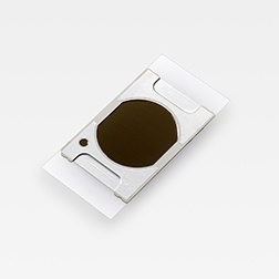
滨松多孔氧化铝制作的辅助离子化基板DIUTHAME(Desorption Ionization Using Through Hole Alumina MEmbrane),可大幅缩减质谱成像分析时待测样品进行离子化所需的前期处理的时间。只要将本产品放置在待测样品上,就能完成质量分析的前期处理。与目前主要的离子化方法之一基质辅助激光解析电离(Matrix-Assisted Laser Desorption/Ionization、下面简称MALDI)方法相比,它将前期处理时间缩短到十分之一。产品具备方便易用、无基质噪声、高重现性、高空间分辨率。本案例为使用辅助离子化基板进行的鼠脑质谱成像。
方案详情

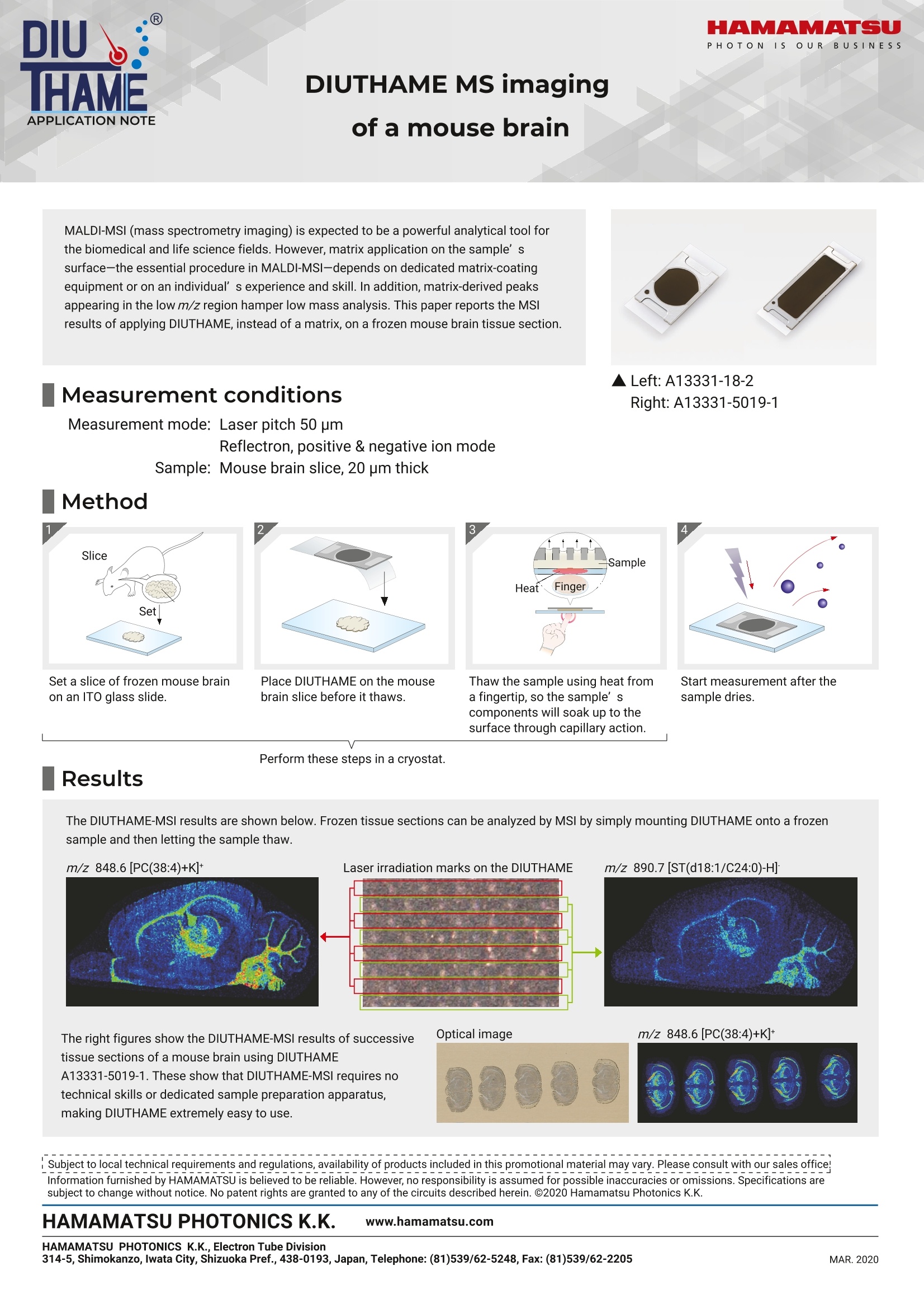
HAMAMATSUPHOTONISs dOUR BUSINESSHAMAMATSU PHOTONICS K.K., Electron Tube Division314-5, Shimokanzo, Iwata City, Shizuoka Pref., 438-0193, Japan, Telephone: (81)539/62-5248, Fax:(81)539/62-2205MAR.2020 DIUTHAME MS imagingof a mouse brain MALDI-MSI (mass spectrometry imaging) is expected to be a powerful analytical tool forthe biomedical and life science fields. However, matrix application on the sample’ssurface-the essential procedure in MALDI-MSI-depends on dedicated matrix-coatingequipment or on an individual’s experience and skill. In addition, matrix-derived peaksappearing in the low m/z region hamper low mass analysis. This paper reports the MSlresults of applying DIUTHAME, instead of a matrix, on a frozen mouse brain tissue section. Measurement conditions ▲Left:A13331-18-2Right: A13331-5019-1 Measurement mode: Laser pitch 50 pmReflectron, positive & negative ion modeSample: Mouse brain slice, 20 pm thick Method 2 4 Set a slice of frozen mouse brainon an ITO glass slide. Place DIUTHAME on the mousebrain slice before it thaws. Start measurement after the sample dries. Perform these steps in a cryostat. Results The DIUTHAME-MSI results are shown below. Frozen tissue sections can be analyzed by MSl by simply mounting DIUTHAME onto a frozensample and then letting the sample thaw. m/z 848.6 [PC(38:4)+K]+Laser irradiation marks on the DIUTHAME m/z 890.7 [ST(d18:1/C24:0)-H] The right figures show the DIUTHAME-MSI results of successivetissue sections of a mouse brain using DIUTHAMEA13331-5019-1. These show that DIUTHAME-MSI requires notechnical skills or dedicated sample preparation apparatus,making DIUTHAME extremely easy to use. Optical image m/z 848.6[PC(38:4)+K]+ 003000003 Subject to local technical requirements and regulations, availability of products included in this promotional material may vary. Please consult with our sales office!Information furnished by HAMAMATSU is believed to be reliable. However, no responsibility is assumed for possible inaccuracies or omissions. Specifications are subject to change without notice. No patent rights are granted to any of the circuits described herein. C2020 Hamamatsu Photonics K.K. HAMAMATSU PHOTONICS K.K. www.hamamatsu.com 滨松多孔氧化铝制作的辅助离子化基板DIUTHAME(Desorption Ionization Using Through Hole Alumina MEmbrane),可大幅缩减质谱成像分析时待测样品进行离子化所需的前期处理的时间。只要将本产品放置在待测样品上,就能完成质量分析的前期处理。与目前主要的离子化方法之一基质辅助激光解析电离(Matrix-Assisted Laser Desorption/Ionization、下面简称MALDI)方法相比,它将前期处理时间缩短到十分之一。产品具备方便易用、无基质噪声、高重现性、高空间分辨率。本案例为使用辅助离子化基板进行的鼠脑质谱成像。
确定
还剩1页未读,是否继续阅读?

产品配置单
滨松光子学商贸(中国)有限公司为您提供《鼠脑切片中质谱成像检测方案(质谱部件)》,该方案主要用于其他中生化检验检测,参考标准--,《鼠脑切片中质谱成像检测方案(质谱部件)》用到的仪器有滨松辅助离子化基板DIUTHAME A13331-18-2
相关方案
更多