


方案详情
文
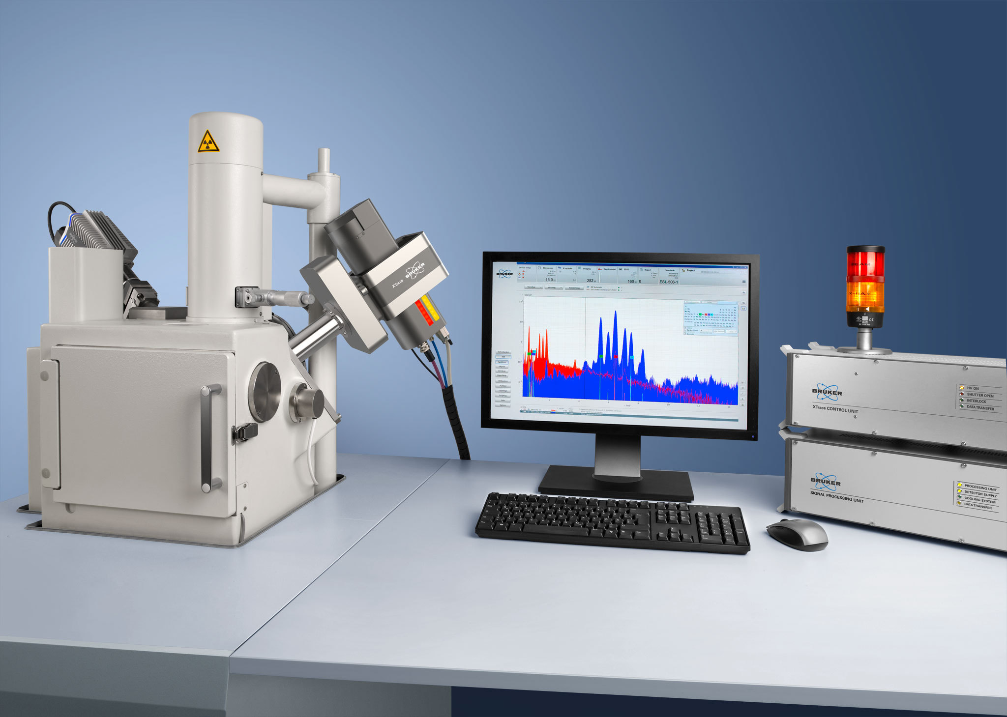
Non-destructive sample analysis with high spatial resolution can be very difficult to accomplish using conventional EDS detectors as sample surface charging, electron beam damage and shading effects by topography are common problems in scanning electron microscopy. The BRUKER XFlash® FlatQUAD silicon drift detector (SDD) overcomes these limitations. The special geometry with an annular arrangement of detector elements offers a very high solid
angle [1]. The XFlash® FlatQUAD detector allows the use of ultra-low beam currents and the investigation of samples with complex topography as demonstrated on two meteorite
fall samples.
方案详情

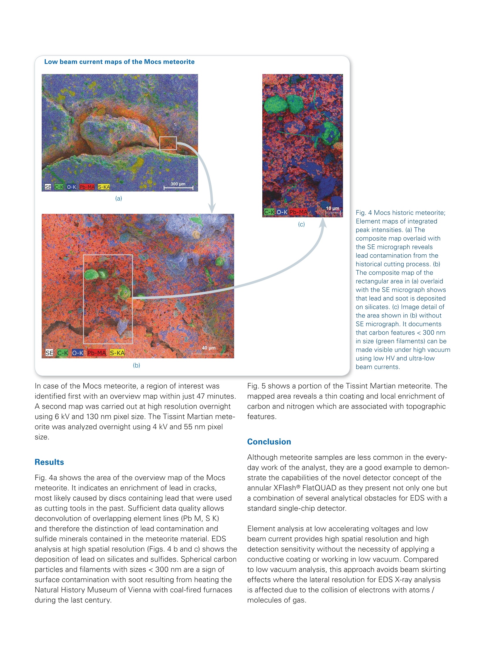
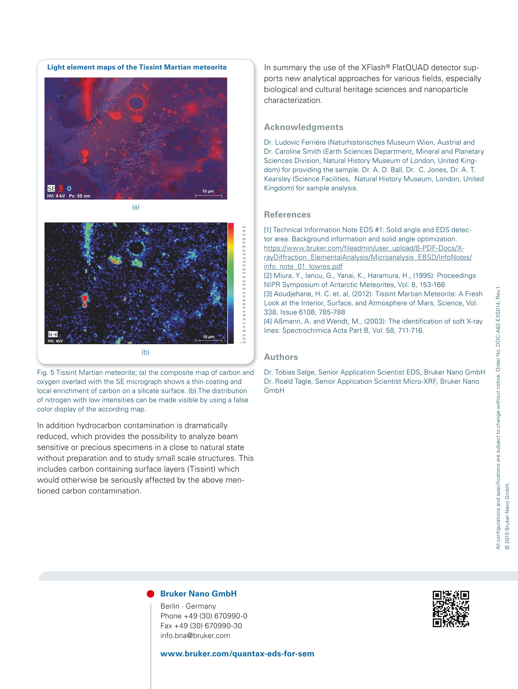
Berlin · Germany Application Note # EDS-12 Application example for theXFlash@ FlatQUAD: High vacuum andlow beam current analysis Non-destructive sample analysis with high spatial resolu-tion can be very difficult to accomplish using conventionalEDS detectors as sample surface charging, electron beamdamage and shading effects by topography are commonproblems in scanning electron microscopy. The BRUKERXFlash@ FlatQUAD silicon drift detector (SDD) overcomesthese limitations. The special geometry with an annulararrangement of detector elements offers a very high solidangle [1]. The XFlash@FlatQUAD detector allows the use ofultra-low beam currents and the investigation of sampleswith complex topography as demonstrated on two meteo-rite fall samples. The Mocs historic meteorite fell on February 2, 1882 in Hun-gary [2]. The historically cut sample (Fig. 1a) was stored atNaturhistorisches Museum Wien (Natural History Museum,Vienna, Austria) for the last century. Studies recently carriedout with the Bruker M4 TORNADO benchtop Micro-XRFspectrometer revealed an unexpected lead enrichmentalong cracks (Fig. 1b). This finding led to a detailed analysisusing the XFlash@ FlatQUAD detector. The second study was carried out on a piece of the Tissint Martian meteoritethat fell in Morocco on July 18, 2011 [3]. Tissint is only thefifth Martian meteorite that people have witnessed fallingto Earth, the last time such an event happened was in1962. Thus, the scientific value of these unique meteoritesexcludes sample preparation. Method The XFlash@FlatQUAD is an annular SDD arrangement which is placed between the pole piece and the sample.This special geometry is ideal for the analysis of topographi-cally complex samples. Shadowing effects are minimized bycollecting X-rays from four separate detector segments withhigh take-off angles. Additionally, the large solid angle of upto 1.1 sr enables EDS analysis with reasonable count ratesusing ultra-low beam currents of <10 pA. As a result, thecharging of nonconductive samples under high vacuum isminimized. This approach therefore enables non-destructiveEDS analysis of sensitive topographic samples,as samplepreparation like polishing and carbon coating is not required. Fig. 1 Mocs historic meteorite; (a) Photograph of the cut sample. The rectangle shows the area analyzed with Micro-XRF. For EDS analysisthe specimen was mounted on a sample holder and analyzed without carbon coating. (b) Video image overlaid with the Micro-XRF map of thelead Lo X-ray line (1472 pixel x676 pixel, 25 um pixel size, 33 min acquisition time) showing lead enrichment along cracks For this study, low accelerating voltages (<6 kV, seealso Tab. 1) were used to examine fine structures. Thisdecreases the interaction volume for electron trajectoriesand the emitted X-rays, as shown in Fig. 2. Table 1 Measurement conditions used for acquiring the mapsdiscussed in this report. Due to the specific geometry of the annularXFlash@ FlatQUAD, the input count rate of the map with low topogra-phy in the crack of the Mocs meteorite is just slightly lower than thatof the overview map. Mocs(overview) Mocs(crack) Tissint Acceleratingvoltage/kV 6 6 4 Beam current/pA <10 <10 <10 Input count rate /kcps 2.9 2.4 1.8 Map resolution/pixels² 800×600 1600x1200 1280x853 Pixel resolution 2 pm 130 nm 55nm Acquisition time 47 min 15h 13h Fig.2 The X-ray excita-tion depth functionsdisplay the intensityof emitted X-raysdepending on theirdepth in the sample.The highest X-rayintensities are emittedmainly from a depth~50 nm for lead M at6 kV (red graph) and~150 nm for carbon Kat 4 kV (blue graph). Itshows that the spatialresolution for EDSX-ray analysis can besignificantly enhancedby using low accele-rating voltages. Depth/nm The peak overlaps of low to intermediate energy X-ray lines(Fig.3) were deconvolved using an extended atomic data-base [4] and an automatic routine. Low beam current maps of the Mocs meteorite (a) 2 10 um C-KKECO-KPb-MA (c) (b) Fig. 4 Mocs historic meteorite;Element maps of integratedpeak intensities.(a) Thecomposite map overlaid withthe SE micrograph revealslead contamination from thehistorical cutting process. (b)The composite map of therectangular area in (a) overlaidwith the SE micrograph showsthat lead and soot is depositedon silicates.(c) Image detail ofthe area shown in (b) withoutSE micrograph. It documentsthat carbon features < 300 nmin size (green filaments) can bemade visible under high vacuumusing low HV and ultra-lowbeam currents. In case of the Mocs meteorite, a region of interest wasidentified first with an overview map within just 47 minutes.A second map was carried out at high resolution overnightusing 6 kV and 130 nm pixel size. The Tissint Martian mete-orite was analyzed overnight using 4 kV and 55 nm pixelsize. Results Fig. 4a shows the area of the overview map of the Mocsmeteorite. It indicates an enrichment of lead in cracks,most likely caused by discs containing lead that were usedas cutting tools in the past. Sufficient data quality allowsdeconvolution of overlapping element lines (Pb M, S K)and therefore the distinction of lead contamination andsulfide minerals contained in the meteorite material. EDSanalysis at high spatial resolution (Figs. 4 b andc) shows thedeposition of lead on silicates and sulfides. Spherical carbonparticles and filaments with sizes < 300 nm are a sign ofsurface contamination with soot resulting from heating theNatural History Museum of Vienna with coal-fired furnacesduring the last century. Fig. 5 shows a portion of the Tissint Martian meteorite. Themapped area reveals a thin coating and local enrichment ofcarbon and nitrogen which are associated with topographicfeatures. Conclusion Although meteorite samples are less common in the every-day work of the analyst, they are a good example to demon-strate the capabilities of the novel detector concept of theannular XFlash@ FlatQUAD as they present not only one buta combination of several analytical obstacles for EDS with astandard single-chip detector. Element analysis at low accelerating voltages and lowbeam current provides high spatial resolution and highdetection sensitivity without the necessity of applying aconductive coating or working in low vacuum. Comparedto low vacuum analysis, this approach avoids beam skirtingeffects where the lateral resolution for EDS X-ray analysisis affected due to the collision of electrons with atoms /molecules of gas. N-Kk 10 pm -HV:4kV (b) Fig. 5 Tissint Martian meteorite; (a) the composite map of carbon andoxygen overlaid with the SE micrograph shows a thin coating andlocal enrichment of carbon on a silicate surface. (b) The distributionof nitrogen with low intensities can be made visible by using a falsecolor display of the according map. In additionhydrocarbon contamination is dramaticallyreduced, which provides the possibility to analyze beamsensitive or precious specimens in a close to natural statewithout preparation and to study small scale structures. Thisincludes carbon containing surface layers (Tissint) whichwould otherwise be seriously affected by the above men-tioned carbon contamination. In summary the use of the XFlash@ FlatQUAD detector sup-ports new analytical approaches for various fields, especiallybiological and cultural heritage sciences and nanoparticlecharacterization. Acknowledgments Dr. Ludovic Ferriere (Naturhistorisches Museum Wien, Austria) andDr. Caroline Smith (Earth Sciences Department, Mineral and PlanetarySciences Division, Natural History Museum of London, United King-dom) for providing the sample. Dr. A. D. Ball, Dr. C. Jones, Dr. A. T.Kearsley (Science Facilities, Natural History Museum, London, UnitedKingdom) for sample analysis. References [1] Technical Information Note EDS #1: Solid angle and EDS detec-tor area. Background information and solid angle optimization.https://www.bruker.com/fileadmin/user upload/8-PDF-Docs/X- rayDiffraction ElementalAnalysis/Microanalysis EBSD/InfoNotes/info note 01 lowres.pdf [2] Miura, Y., lancu, G., Yanai, K., Haramura, H., (1995): Proceedings NIPR Symposium of Antarctic Meteorites, Vol. 8,153-166 [3] Aoudjehane, H. C. et. al, (2012): Tissint Martian Meteorite: A Fresh Look at the Interior, Surface, and Atmosphere of Mars, Science, Vol.338, Issue 6108, 785-788 [4] ABmann, A. and Wendt, M., (2003): The identification of soft X-raylines: Spectrochimica Acta Part B, Vol.58,711-716. Authors Dr. Tobias Salge, Senior Application Scientist EDS, Bruker Nano GmbHDr. Roald Tagle, Senior Application Scientist Micro-XRF, Bruker NanoGmbH The Mocs historic meteorite fell on February 2, 1882 in Hungary [2]. The historically cut sample (Fig. 1a) was stored at Naturhistorisches Museum Wien (Natural History Museum, Vienna, Austria) for the last century. Studies recently carried out with the Bruker M4 TORNADO benchtop Micro-XRF spectrometer revealed an unexpected lead enrichment along cracks (Fig. 1b). This finding led to a detailed analysis using the XFlash® FlatQUAD detector. The second study was carried out on a piece of the Tissint Martian meteorite that fell in Morocco on July 18, 2011 [3]. Tissint is only the fifth Martian meteorite that people have witnessed falling to Earth, the last time such an event happened was in 1962. Thus, the scientific value of these unique meteorites excludes sample preparation.
确定

还剩2页未读,是否继续阅读?

产品配置单

布鲁克电子显微纳米分析仪器部为您提供《陨石中元素含量检测方案(电镜部件)》,该方案主要用于非金属矿产中痕量元素检测,参考标准--,《陨石中元素含量检测方案(电镜部件)》用到的仪器有布鲁克 QUANTAX 微区荧光光谱仪、布鲁克 平插式能谱仪FlatQUAD、布鲁克 ESPRIT 四合一分析软件
相关方案
更多