


方案详情
文
本方案主要使用Incucyte®活细胞分析系统和Incucyte®类器官分析软件,对类器官的生长、数目和形态进行动态监测和定量分析。
方案详情

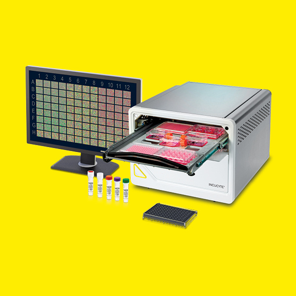
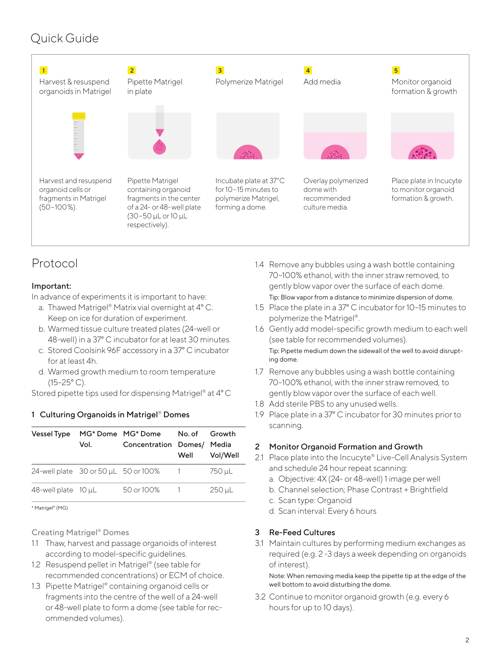
SARTORIUS2 Incucyte@Organoid Culture QC For the Quantification of Organoid Growth inMatrigel Domes This protocol provides an overview for culturing,monitoringand measuring organoid growth, counts and morphologyof Brightfield images in Matrigeldomes utilizing theIncucyte@ Live-Cell Analysis System and Incucyte°Organoid Analysis Software. General Guidelines ▪ Follow manufactures guidelines for thawing and storingof 100% Matrigel.Thaw Corning@Matrigelovernight bysubmerging the vial in ice cold water in the rear of arefrigerator (4°C). Do not allow Matrigel to warm toroom temperature at any time as this will inducepolymerization. ▪ Following dome creation (in 50-100% Matrigel) andmedia addition, remove bubbles from dome or wells re-spectively by gently squeezing a wash bottle containing70-100% ethanol, with the inner straw removed, to blowvapor over the surface of each well. After organoid seeding and all media changes,placethe plate in the Incucyte Live-Cell Analysis System andallow the plate to warm to 37°C for 30 minutes prior toscanning. Required Materials 24-well or 48-well flat bottom TC-treated microplate(Corning Cat. No.3526,Cat. No. 3548respectively) · Matrigel Growth Factor Reduced (GFR), Phenol Red-Free (Corning Cat. No.356231) Organoids of interest Organoid Specific Growth Medium ■ Manual single-channel pipettes ■Wet Ice Incucyte°Organoid Analysis Software Module, version2020B (Cat. No. 9600-0034-A00) Optional Materials ■ IncucyteCoolsink 96F (Cat. No.1500-0080) ·StemCell Technologies, Mouse Intestinal Organoids Cat.No. 70931; Mouse Pancreatic Organoids Cat. No.70933;Mouse Hepatic Organoids Cat.No.70932 IntestiCultMOGM Cat.No.06005;PancreaCultOGMCat. No.06040;HepatiCult'MOGM Cat.No.06030 Protocol Important: In advance of experiments it is important to have: a. Thawed Matrigel Matrix vial overnight at 4°C.Keep on ice for duration of experiment. b. Warmed tissue culture treated plates (24-well or48-well) in a 37°C incubator for at least 30 minutes. s. Stored Coolsink 96F accessory in a 37°C incubatorfor at least 4h. d. Warmed growth medium to room temperature(15-25°C). Stored pipette tips used for dispensing Matrigel at 4°C 1 Culturing Organoids in Matrigel Domes Vessel Type MG*Dome MG*Dome No. of Growth Vol. Concentration Domes/1 Media Well Vol/Well 24-wellplate 30 or 50 uL 50 or100% 750 uL 48-well plate 10uL e 50 or100% 250 uL Creating Matrigel°Domes 1.1 Thaw, harvest and passage organoids of interestaccording to model-specific guidelines. 1.2 RFesuspend pellet in Matrigel@ (see table forrecommended concentrations) or ECM of choice. 1.3 PFipette Matrigel°containing organoid cells orfragments into the centre of the well of a 24-wellor 48-well plate to form a dome (see table for rec-ommended volumes). 1.4RRemove any bubbles using a wash bottle containing70-100% ethanol, with the inner straw removed,togently blow vapor over the surface of each dome. Tip: Blow vapor from a distance to minimize dispersion of dome. 1.5PPlace the plate in a 37°C incubator for 10-15 minutes topolymerize the Matrigel. 1.6Gently add model-specific growth medium to each well(see table for recommended volumes).Tip: Pipette medium down the sidewall of the well to avoid disrupt-ing dome. 1.7Remove any bubbles using a wash bottle containing70-100% ethanol, with the inner straw removed, togently blow vapor over the surface of each well. 1.8AAdd sterile PBS to any unused wells. 1.9PPlace plate in a 37°C incubator for 30 minutes prior toscanning. 2NMonitor Organoid Formation and Growth 2.1Place plate into the Incucyte Live-Cell Analysis Systemand schedule 24 hour repeat scanning: a. Objective: 4X (24-or 48-well) 1 image per wellb. Channel selection; Phase Contrast + Brightfield c. Scan type:Organoid d. Scan interval: Every 6 hours 3 Re-Feed Cultures 3.1Maintain cultures by performing medium exchanges asrequired (e.g. 2-3 days a week depending on organoidsof interest).Note: When removing media keep the pipette tip at the edge of the well bottom to avoid disturbing the dome. 3.2 Continue to monitor organoid growth (e.g. every6hours for up to 10 days). .Analysis Guidelines 1 Create a New Analysis Definition 2 Data Interpretation a. In the Analysis Wizard window select'Organoid'Analysis lype. Once the Analysis Job is complete the following primarymetrics are provided: b. Select a set of representative images. a. Organoid Object Count. This metric represents thenumber of objects per image (well). c. Adjust the Background/cells slider to determine theboundary of the organoidobjects d. Evaluate the Brightfield (BF) mask and refine filterparameters accordingly.'Preview All’ to ensure param-eters set appropriately mask all representative imageswithin the collection. b. Organoid Object Total Area.This metric represents thetotal area of BF objects within the image (well) and isrecommended for tracking organoid size over time. c. Organoid Object Average Eccentricity. This metricrepresents how round the organoids are. e. Adjust the Edge split slider to delineate betweenindividual organoid objects. d. Organoid Darkness. This metric is available for trackingchanges in organoid brightness over time. f.Evaluate the BF mask and refine filter parametersaccordingly.Preview All' to ensure parameters setappropriately mask all representative images withinthe collection. g. Once satisfied with all parameters, complete theLaunch Wizard analysis by selecting the scan timesand wells to be analyzed. Note: If your experiment is in progress you will have an option to checkAnalyze Future Scans'to perform real-time analysis. For Research Use Only. Not For Therapeutic or Diagnostic Use. Sales and Service North America APAC Essen BioScience Inc. Essen BioScience K.K. Contacts 300 West Morgan Road 4th Floor Daiwa Shinagawa North Ann Arbor, Michigan, 48108 Bldg. USA 1-8-11 Kita-Shinagawa Telephone +1734 769 1600 Shinagawa-ku, Tokyo For further contacts, visit E-Mail:orders.USO7@sartorius.com 140-0001 Www.sartorius.com Japan Europe Telephone: +81 3 6478 5202 Essen BioScience Ltd. E-Mail:orders.USO7@sartorius.com Units 2 & 3 The Quadrant Essen BioScience, A Sartorius Company Newark Close www.sartorius.com/incucyte Royston Hertfordshire E-Mail: AskAScientist@sartorius.com SG8 5HL United Kingdom Telephone +44 1763 227400 Specifications subject to change without notice. E-Mail: ◎2020. All rights reserved. Incucyte, Essen BioScience, and all names of Essen BioScience euorders.UK03@sartorius.com products are registered trademarks and the property of Essen BioScience unless otherwise Status:06|2020 肠道类器官作为肠道的三维细胞培养模型,已经用于证明冠状病毒SARS-CoV-2可以感染肠道细胞并繁殖。这些模型表明,SARS-CoV-2也可能在人体肠壁细胞中感染和复制。甚至有可能如研究人员所怀疑的那样,人体肠道是SARS-CoV-2传播的重要途径。也就是说,SARS-CoV-2通过胃肠道器官以及呼吸器官的传播,可能是COVID-19强有力破坏性扩散的原因。从患者分离的SARS-CoV-2病毒颗粒的电镜图COVID-19患者表现出与呼吸器官有关的各种症状(如咳嗽、打喷嚏、呼吸急促和发烧),疾病通过微小的飞沫传播,而飞沫主要通过咳嗽和打喷嚏扩散。然而,三分之一的患者也有胃肠道症状,如恶心和腹泻。此外,在呼吸道症状消失很长一段时间后,仍可在人的粪便中检测到这种病毒。这表明病毒也可以通过所谓的粪口传播扩散。虽然呼吸器官和胃肠器官看起来很不一样,但它们有一些关键的相似之处。一个特别有趣的相似之处是都存在ACE2受体,引发COVID-19的SARS-CoV-2病毒通过该受体进入细胞。肠道内部含有ACE2受体。肠道类器官,右侧感染了冠状病毒sars-cov-2。冠状病毒呈白色,类器官本身呈蓝色和绿色。然而,目前还不清楚肠道细胞是否会被感染并产生病毒颗粒。为了探索肠道途径的可能性,荷兰的科学家通过原代肠上皮干细胞培养生成了人类小肠类器官(hSIOs)。荷兰皇家科学院干细胞与发育生物学研究所、伊拉斯谟大学医学中心和马斯特里赫特大学的科学家代表宣称,这类hSIOs包含了体内上皮的所有增殖和分化细胞类型。当在四种不同的培养条件下生长时,hSIOs含有不同ACE2表达量的细胞,因此理论上可以被SARS-CoV-2感染。为了观察细胞是否真的被感染,科学家们对其进行了密切的观察,如5月1日发表在《科学》杂志上的一篇文章(《SARS-CoV-2有效地感染人类肠道肠上皮细胞》)所述。文章作者写道:“在共聚焦显微镜和电子显微镜下证实,在hSIOs中,肠上皮细胞很容易被SARS-CoV和SARS-CoV-2感染。”“因此,检测到显著的传染性病毒颗粒滴度。mRNA表达分析显示了一个通用病毒反应程序的强烈诱导。”基本上,科学家们发现病毒同时感染了成熟的肠上皮细胞和前体肠上皮细胞,这些细胞是排列在肠道内表面的肠吸收上皮细胞。这张扫描电子显微镜图片显示SARS-CoV-2(橙色)从实验室培养的细胞(绿色)表面出现。科学家们还发现,这种病毒激发了与抗病毒反应有关的基因活性。值得注意的是,所有类器官模型的感染率是相似的,这表明即使少量的ACE2也足以让病毒感染上皮细胞。本研究的通讯作者、伊拉斯谟大学医学中心的研究人员Bart Haagmans博士说:“这项研究中的观察结果提供了明确的证据,证明SARS-CoV-2可以在胃肠道细胞中繁殖。”“然而,我们目前还不知道存在于COVID-19患者肠道中的SARS-CoV-2是否在传播中起重要作用。我们的研究结果表明,我们应该更密切地观察这种可能性。”本研究与其他最近的研究一致,这些研究发现大部分COVID-19患者存在胃肠道症状,而且在无呼吸道症状患者的粪便中也发现病毒。我们可能需要特别关注有胃肠道症状的患者。因此,可能不仅需要使用鼻咽拭子,还需要使用直肠拭子或粪便样本进行更广泛的检测。与此同时,研究人员正在继续合作,以进一步研究COVID-19。他们正在通过对比SARS-CoV-2感染的肺和肠道类器官来研究肺部感染和肠道感染之间的差异。本方案主要使用Incucyte®活细胞分析系统和Incucyte®类器官分析软件,对类器官的生长、数目和形态进行动态监测和定量分析。点击下载Protocol了解详细实验步骤
确定

还剩1页未读,是否继续阅读?

产品配置单

德国赛多利斯集团为您提供《粪便中病毒检测方案(高内涵成像)》,该方案主要用于粪便中生化检验检测,参考标准--,《粪便中病毒检测方案(高内涵成像)》用到的仪器有赛多利斯 Incucyte® SX5 活细胞分析仪
该厂商其他方案
更多