


方案详情
文
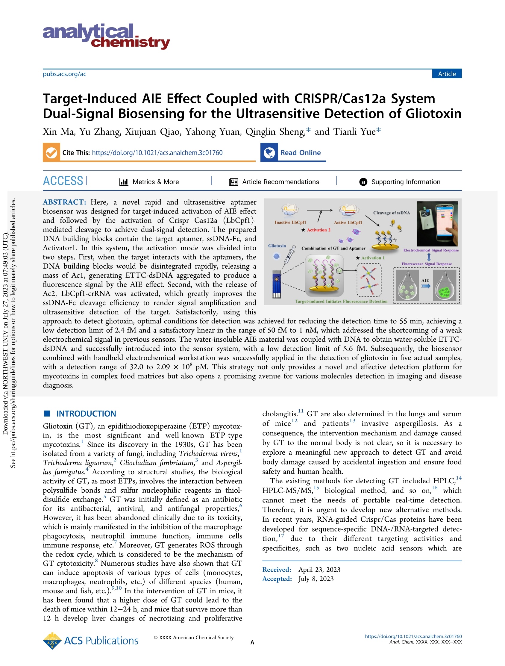
一种新型的快速、超灵敏的电化学生物传感器,用于靶向诱导激活AIE效应和Crispr Cas12a (LbCpf1)的无差别剪切功能,实现双信号检测胶霉毒素。构建的DNA传感单元包含适配体、ssDNA-Fc和Activator1。在本系统中,激活模式分为两个步骤。首先,当靶标与适配体相互作用时,DNA传感单元迅速分解启动链转移反应,释放出大量Ac1,通过AIE效应聚集ETTC-dsDNA产生荧光信号。其次,ETTC-dsDNA在聚集过程中释放Ac2,激活LbCpf1的无差别剪切功能,极大地提高了ssDNA-Fc的剪切效率,实现了体系的信号放大和对靶标的超灵敏检测。利用该方法检测胶霉毒素,电化学信号检测限低至2.4 fM,在50 fM~1 nM范围内具有良好的线性关系,检测时间缩短至55 min,解决了以往传感器电化学信号弱的缺点。同时将不溶于水的AIE材料与DNA偶联得到水溶性ETTC-dsDNA,并成功引入水介质传感系统,作为荧光响应信号,检测限低至5.6 fM。研究结果表明,通过结合手持式电化学工作站,该传感器成功应用于5种实际样品中的胶霉毒素的检测,检测范围可达到32.0~2.09×10⁸ pM。该方法不仅为复杂食物基质中真菌毒素的检测提供了一种新颖有效的检测平台,而且为分子成像和疾病诊断领域开辟了一条有前景的途径。
方案详情

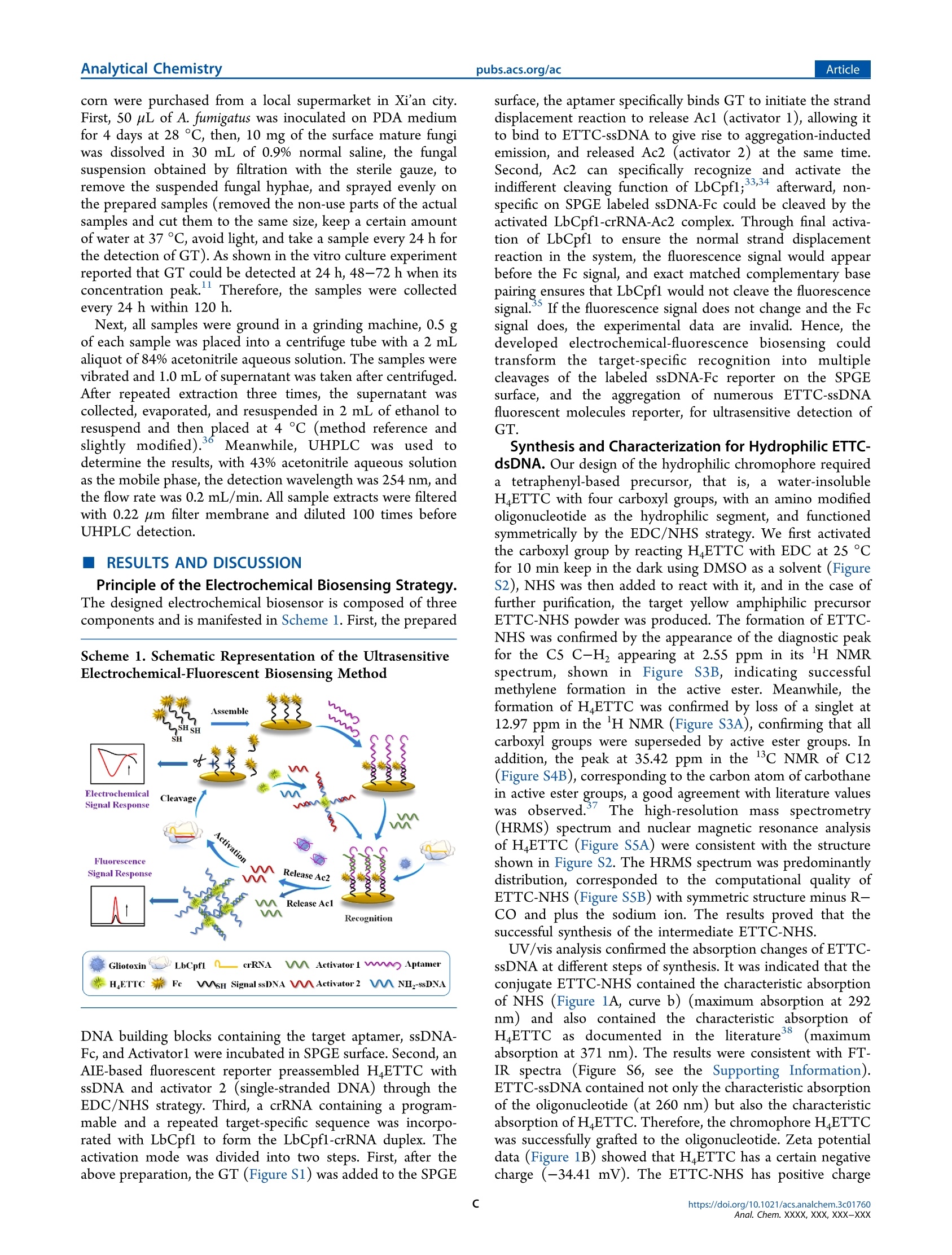
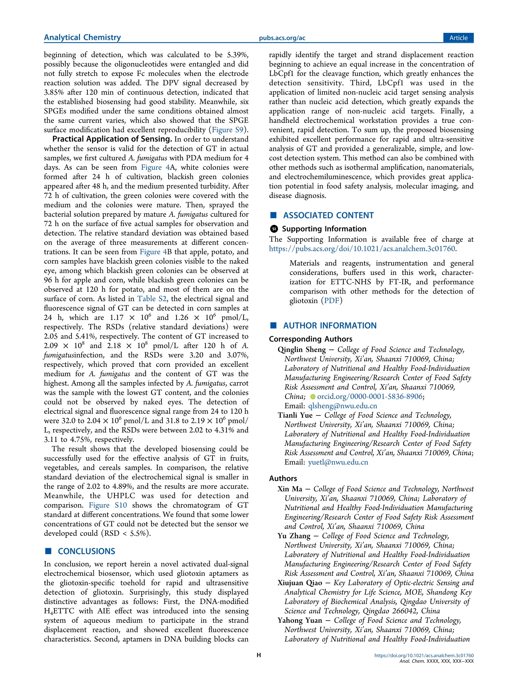
本文提出了一种新型的快速、超灵敏的电化学生物传感器,用于靶向诱导激活AIE效应和Crispr Cas12a (LbCpf1)的无差别剪切功能,实现双信号检测胶霉毒素。构建的DNA传感单元包含适配体、ssDNA-Fc和Activator1。在本系统中,激活模式分为两个步骤。首先,当靶标与适配体相互作用时,DNA传感单元迅速分解启动链转移反应,释放出大量Ac1,通过AIE效应聚集ETTC-dsDNA产生荧光信号。其次,ETTC-dsDNA在聚集过程中释放Ac2,激活LbCpf1的无差别剪切功能,极大地提高了ssDNA-Fc的剪切效率,实现了体系的信号放大和对靶标的超灵敏检测。利用该方法检测胶霉毒素,电化学信号检测限低至2.4 fM,在50 fM~1 nM范围内具有良好的线性关系,检测时间缩短至55 min,解决了以往传感器电化学信号弱的缺点。同时将不溶于水的AIE材料与DNA偶联得到水溶性ETTC-dsDNA,并成功引入水介质传感系统,作为荧光响应信号,检测限低至5.6 fM。研究结果表明,通过结合手持式电化学工作站,该传感器成功应用于5种实际样品中的胶霉毒素的检测,检测范围可达到32.0~2.09×10⁸ pM。该方法不仅为复杂食物基质中真菌毒素的检测提供了一种新颖有效的检测平台,而且为分子成像和疾病诊断领域开辟了一条有前景的途径。原文链接:https://doi.org/10.1021/acs.analchem.3c01760本文中进行电化学测试的仪器为荷兰PalmSens,型号:EmStat3Blue便携式电化学分析仪,由雷迪美特中国有限公司提供。analyticanistryArticle AnalyticalChemistryArticlepubs.acs.org/ac pubs.acs.org/ac Target-InducedAIEEfefctCoupledwithCRISPR/Cas12aSystemDual-SignalBiosensingfortheUltrasensitiveDetectionofGliotoxin XinMa,YuZhang,XiujuanQiao,YahongYuan,QinglinSheng,*andTianliYue* CiteThis:https://doi.org/10.1021/acs.analchem.3c01760 ReadOnline ACCESS dil M M e e t t r r i i c c s s & More & More A A r r t t i i c c l l e e R R e e c c o o m m m m e e n n d d a a t t i i o o n n s s GD S S u u p p p p o o r r t t i in ng g I I n n f f o o r r m m a a t t i i o o n n INTRODUCTION daiagnosis.Gliotoxin(GT),anepidithiodioxopiperazine(ETP)mycotox-in,isthemostsignificantandwell-knownETP-type mycotoxins.1Sinceitsdiscoveryinthe1930s,GThasbeenisolatedfromavarietyoffungi,includingTrichodermavirens,1Trichodermalignorum,2Gliocladiumfimbriatum,3andAspergil-lusfumigatus.4Accordingtostructuralstudies,thebiological activityofGT,asmostETPs,involvestheinteractionbetweenpolysulfidebondsandsulfurnucleophilicreagentsinthiol-disulfideexchange.5GTwasinitiallydefinedasanantibioticforitsantibacterial,antiviral,andantifungalproperties,6However,ithasbeenabandonedclinicallyduetoitstoxicity,whichismainlymanifestedintheinhibitionofthemacrophage phagocytosis,neutrophilimmunefunction,immunecellsimmuneresponse,etc.7Moreover,GTgeneratesROSthrough theredoxcycle,whichisconsideredtobethemechanismofGTcytotoxicity.8NumerousstudieshavealsoshownthatGT caninduceapoptosisofvarioustypesofcells(monocytes,macrophages,neutrophils,etc.)ofdifefrentspecies(human,mouseandfsih,etc.).9,10IntheinterventionofGTinmice,ithasbeenfoundthatahigherdoseofGTcouldleadtothe deathofmicewithin12−24h,andmicethatsurvivemorethan12hdevelopliverchangesofnecrotizingandproliferative cholangitis.11GTarealsodeterminedinthelungsandserumofmice12andpatients13invasiveaspergillosis.Asa consequence,theinterventionmechanismanddamagecausedbyGTtothenormalbodyisnotclear,soitisnecessaryto exploreameaningfulnewapproachtodetectGTandavoidbodydamagecausedbyaccidentalingestionandensurefood safetyandhumanhealth. TheexistingmethodsfordetectingGTincludedHPLC,14HPLC-MS/MS,15biologicalmethod,andsoon,16which cannotmeettheneedsofportablereal-timedetection.Therefore,itisurgenttodevelopnewalternativemethods.Inrecentyears,RNA-guidedCrispr/Casproteinshavebeendevelopedforsequence-specifciDNA-/RNA-targeteddetec-tion,17duetotheirdifferenttargetingactivitiesandspecifciities,suchastwonucleicacidsensorswhichare Received: April23,2023 Accepted:July8,2023 basedonDETECTR(DNAendonuclease-targetedCRISPRtransreporter)andSHERLOCK(specifcihigh-sensitivity enzymaticreporterunlocking).18,19BiosensorsbasedontheCrispr/Cassystemhavebeensuccessfullyappliedtothe detectionofmiRNAspecifciityandsensitivityincomplexbiologicalsamplessuchasserums,tissuesandcellextracts,etc.20CRISPR-cas12a(Cpf1)hasbeenreportedtocleavethetargetDNA,aswellasindiscriminatelycleaveanynon-target ssDNA.21Interestingly,thecleavageofnon-targetssDNAcansignifciantlyimprovethesensitivityofdetectioninmany applications.Xiongetal.22exploitedthepresenceoftargetstoinduceCrisprCas12aactivationandquantitativedetectionof ATPandNa+.Wangetal.23developedamethodforsingle-moleculecountingofFTOinhumancancercellsandbreast tissuesbyusingrollingcircletranscription(RCT)todrivethereactionofCrisprCas12a.Alongwiththedevelopmentof activatableCRISPRsensors,electrochemistryhasattractedextensiveattentionforitsadvantageofcost-efefctiveness,microminiaturization,fastreactionspeed,andhighsensitivity.Inrecentyears,auniversalelectrochemicalbiosensorbasedon CRISPRhasbeenappliedforthemeasurementofproteinandDNA.Zhangetal.24reportedtheconstructionofelectro-chemicalsensorsforlinearDNAandhairpinDNAusingRNA-guidedCrisprCas12aproteinforthehighlysensitivedetection oftargetmoleculesHPV-16andHPV-18incomplexmatrices.Insensinganalysis,efcfieintandnovelfluorescentmaterialsare alsofrequentlyusedstrategiesforthedetectionofmycotoxinsouttheotherside.Highlyefcfieintluminescentmolecular materialshavebeenhighlyconcernedbyresearchersbecausetheyhaveimportantapplicationvaluesinmanyfeilds,suchas luminescentdevices,chemicalsensors,biologicalprobes,etc.25,26In2001,Tangetal.27fristreportedtheaggregation-inducedemission(AIE)efefct.AIEmoleculeshavebeenwidelyusedinthefeildofchemicalbiosensors,protein conformation,andchiralrecognition.Forinstance,Qinetal.28synthesizedatraceableself-assembledmicelle(TPR@DOX)withAIEcharacteristicsforhigh-qualityimaging.Inaddition,ithasbeenreportedthathigh-fidelityandefcfieincynucleicacid couldbeintroducedintoAIEprobes,andthedetectionofaspecifciDNAwasrealizedthroughspecifciornon-specifci binding.29,30Forinstance,Tangetal.30combinedbisazide-functionalizedfluorescein(AIEgen)withssDNAtoproducea double-armedAIEprobethathybridizedwithDNAtolimitthefreerotationofthecombinedAIEgentoenhancesignal output. SincetheinteractionbetweenAIEfluoresceinandbiomoleculesaremainlyhydrophobicandelectrostatic,very poorornon-selectiveofAIEfluoresceintobiologicaltargets.AspecialexampleisthatcationicTPEderivativescanrespondto difefrentG-quadruplexsequenceswhenformingadoublechainwithcomplementaryDNAchains.28,31Thedetectionof non-nucleic-acidmoleculeshasalsobeenreportedrecentlyabouttheCrispr/Cassystem.32However,comparedwitha largenumberofPOCsensors,thesmallamountoftargetresponseswouldleadtolimitedtargetsdetection.Themain challengeindevelopingofsensorsfornon-nucleic-acidtargetsistofindanapproachtoconvertthetargetrecognitionevents intoacleavageactivityefefctoftheCasproteins,usuallyactivatedbyspecifcibindingofthecrRNAtomatchedDNA (hereinafterreferredtoasthe“activator”),facilitatingtheconstructionofsimpleandsensitivebiosensors.33Wereport hereageneraldesignstrategyofanactivatedCRISPR-Cassensorthatutilizesthespecifciaptamerrecognitionofthe targetsbasedonthenon-nucleic-acidtargetresponsesfunctionofDNAbuildingblocks.UsingthessDNA-Fcreporterfroma handheldelectrochemicalworkstationandthehighturnoverLbCpf1ssDNaseactivity,significantchangesinelectro-chemicalandfluorescenceresponseswereachievedsimulta-neously.Underoptimalconditions,targetmoleculesaslowas 50fMweredetectedinabout55min,withalinearrangefrom50fMto1nM.Theestablishedbiosensingcouldbeappliedto thedetectionoftargetmoleculesincomplexsamples,withexcellentsensitivity,specifciity,andeasyimplementation. EXPERIMENTALSECTION SynthesisofNovelTPE-BasedFluorescentMaterialETTC-dsDNA.H4ETTC(10.15mg,0.012mM)andEDC(10mg,0.052mM)weremixedinDMSO(500mL)toavoidlightoscillated10min.NHS(5mg,0.043mM)wasaddedtothe mixtureandoscillatedwithoutlightfor48hatnormaltemperature.Then,thecrudeproductwaspurifeidonasilica gelcolumnusingEtOAc/cyclohexane(5.5:1)aseluent.Aftervacuumdrying,ETTC-NHSwasobtainedasayellowpowder.NH2-ssDNA(500nM)wasdissolvedinNaHCO3(0.1M,250nL)aqueoussolution,andETTC-NHS(100nM)was dissolvedinDMSO(250 L).Thetwosolutionswereintensivemixedandincubatedat37°Cfor24hawayfrom light.Themixturewaspurifeidandconcentratedwithanultraflitrationtube,and10mMTris−EDTAsolutionwas addedtoobtainthedesiredconcentrationoftheoriginalsolutionandstoredat4°C.Finally,ETTC-ssDNA(20nL)wasmixedwithActivator2(20TL,100nM)solution(dissolvedin10mMTris−EDTA)andpreparedintoETTC-dsDNAsolution(50nM)andstoredat4°C. PreparationoftheDNABuildingBlockBiosensingElectrode.Forthemodifciation,theSPGEswerewashed thoroughlyandthenactivatedinH2SO4(0.5M)solution.BeforeimmobilizationontheSPGEsurface,thiolatedssDNA-Fcwasdissolvedin100mMTris−HClbufefr.Then,10sLofssDNA-Fc(100nM)wasgentlyaddedtothesurfaceofSPGE andincubatedfor16hat4°C,rinsedandthengentlyadded10eLofMCH(1mM)toblockthenon-specifcisites,resultinginagoodarrangedDNAmonolayer.Next,7tLofaptamer(100nM)solutionwasgentlyaddedtotheSPGE surface,andincubatedfor4hat4°C.Finally,7SLofActivator1(100nM)wasgentlyaddedtothesurfaceofthe SPGE,incubatedfor4hat4°C.UponcompletionofallDNAmodifciations,theSPGEwasthoroughlyrinsedanduseditfor thefollowingexperimentaloperations. GliotoxinDetectionProtocol.BeforedetectionofGT,6lLofpreparedLbCpf1/crRNAsolution(55nMLbCpf1and 45nMcrRNAwerereactedawaythelightat25°Cfor30min)wasmixedwith2eLofETTC-dsDNAand2rLof gliotoxinsolutions.Then,10LLofthemixturesolutionwasaddedtothemodifeidbiosensorplatformsurfacedropwiseand incubatedfor1hat37°CtoconductLbCpf1-mediatedcleavageexperiments.Afterward,thesolutionontheSPGE surfacewascollectedanddilutedto100iLwith10mMTris−EDTAbufefrforfluorescencemeasurement.Subsequently,the conditionsofelectrochemicaldifefrentialpulsevoltammetry(DPV)measurementswereasfollows,thepotentialscanning speedwas0.1V/s,theintervaltimewas0.02s,thesteppotentialwas10mV,thepulsepotentialwas0.2V,andthe scanningrangewas0.65~0.1V. PracticalSamplesPretreatmentandDetection.Theactualsamplesincludingapple,carrot,sweetpotato,potatoand cornwerepurchasedfromalocalsupermarketinXi’ancity.First,50hLofA.fumigatuswasinoculatedonPDAmedium for4daysat28°C,then,10mgofthesurfacematurefungiwasdissolvedin30mLof0.9%normalsaline,thefungal suspensionobtainedbyflitrationwiththesterilegauze,toremovethesuspendedfungalhyphae,andsprayedevenlyon thepreparedsamples(removedthenon-usepartsoftheactualsamplesandcutthemtothesamesize,keepacertainamount ofwaterat37°C,avoidlight,andtakeasampleevery24hforthedetectionofGT).Asshowninthevitrocultureexperiment reportedthatGTcouldbedetectedat24h,48−72hwhenitsconcentrationpeak.11Therefore,thesampleswerecollected every24hwithin120h. Next,allsamplesweregroundinagrindingmachine,0.5gofeachsamplewasplacedintoacentrifugetubewitha2mL aliquotof84%acetonitrileaqueoussolution.Thesampleswerevibratedand1.0mLofsupernatantwastakenaftercentrifuged.Afterrepeatedextractionthreetimes,thesupernatantwascollected,evaporated,andresuspendedin2mLofethanolto resuspendandthenplacedat4°C(methodreferenceandslightlymodified).36Meanwhile,UHPLCwasusedto determinetheresults,with43%acetonitrileaqueoussolutionasthemobilephase,thedetectionwavelengthwas254nm,and theflowratewas0.2mL/min.Allsampleextractswerefliteredwith0.22omflitermembraneanddiluted100timesbefore UHPLCdetection. RESULTSANDDISCUSSION PrincipleoftheElectrochemicalBiosensingStrategy.Thedesignedelectrochemicalbiosensoriscomposedofthree componentsandismanifestedinScheme1.First,theprepared Scheme1.SchematicRepresentationoftheUltrasensitiveElectrochemical-FluorescentBiosensingMethod DNAbuildingblockscontainingthetargetaptamer,ssDNA-Fc,andActivator1wereincubatedinSPGEsurface.Second,an AIE-basedfluorescentreporterpreassembledH4ETTCwithssDNAandactivator2(single-strandedDNA)throughthe EDC/NHSstrategy.Third,acrRNAcontainingaprogram-mableandarepeatedtarget-specifcisequencewasincorpo-ratedwithLbCpf1toformtheLbCpf1-crRNAduplex.Theactivationmodewasdividedintotwosteps.First,afterthe abovepreparation,theGT(FigureS1)wasaddedtotheSPGE surface,theaptamerspecifciallybindsGTtoinitiatethestranddisplacementreactiontoreleaseAc1(activator1),allowingit tobindtoETTC-ssDNAtogiverisetoaggregation-inductedemission,andreleasedAc2(activator2)atthesametime.Second,Ac2canspecificallyrecognizeandactivatetheindifefrentcleavingfunctionofLbCpf1;33,34afterward,non-specifcionSPGElabeledssDNA-FccouldbecleavedbytheactivatedLbCpf1-crRNA-Ac2complex.Throughfinalactiva-tionofLbCpf1toensurethenormalstranddisplacementreactioninthesystem,thefluorescencesignalwouldappear beforetheFcsignal,andexactmatchedcomplementarybasepairingensuresthatLbCpf1wouldnotcleavethefluorescence signal.35IfthefluorescencesignaldoesnotchangeandtheFcsignaldoes,theexperimentaldataareinvalid.Hence,the developedelectrochemical-fluorescencebiosensingcouldtransformthetarget-specificrecognitionintomultiple cleavagesofthelabeledssDNA-FcreporterontheSPGEsurface,andtheaggregationofnumerousETTC-ssDNA fluorescentmoleculesreporter,forultrasensitivedetectionof GTS.ynthesisandCharacterizationforHydrophilicETTC- dsDNA.Ourdesignofthehydrophilicchromophorerequiredatetraphenyl-basedprecursor,thatis,awater-insoluble H4ETTCwithfourcarboxylgroups,withanaminomodifeidoligonucleotideasthehydrophilicsegment,andfunctioned symmetricallybytheEDC/NHSstrategy.WefristactivatedthecarboxylgroupbyreactingH4ETTCwithEDCat25°C for10minkeepinthedarkusingDMSOasasolvent(FigureS2),NHSwasthenaddedtoreactwithit,andinthecaseof furtherpurifciation,thetargetyellowamphiphilicprecursorETTC-NHSpowderwasproduced.TheformationofETTC-NHSwasconfrimedbytheappearanceofthediagnosticpeakfortheC5C−H2appearingat2.55ppminits1HNMR spectrum,showninFigureS3B,indicatingsuccessfulmethyleneformationintheactiveester.Meanwhile,the formationofH4ETTCwasconfrimedbylossofasingletat12.97ppminthe1HNMR(FigureS3A),confrimingthatall carboxylgroupsweresupersededbyactiveestergroups.Inaddition,thepeakat35.42ppminthe13CNMRofC12(FigureS4B),correspondingtothecarbonatomofcarbothaneinactiveestergroups,agoodagreementwithliteraturevalues wasobserved.37Thehigh-resolutionmassspectrometry(HRMS)spectrumandnuclearmagneticresonanceanalysis ofH4ETTC(FigureS5A)wereconsistentwiththestructureshowninFigureS2.TheHRMSspectrumwaspredominantly distribution,correspondedtothecomputationalqualityofETTC-NHS(FigureS5B)withsymmetricstructureminusR−COandplusthesodiumion.TheresultsprovedthatthesuccessfulsynthesisoftheintermediateETTC-NHS. UV/visanalysisconfrimedtheabsorptionchangesofETTC-ssDNAatdifefrentstepsofsynthesis.Itwasindicatedthatthe conjugateETTC-NHScontainedthecharacteristicabsorptionofNHS(Figure1A,curveb)(maximumabsorptionat292nm)andalsocontainedthecharacteristicabsorptionofH4ETTCasdocumentedintheliterature38(maximum absorptionat371nm).TheresultswereconsistentwithFT-IRspectra(FigureS6,seetheSupportingInformation).ETTC-ssDNAcontainednotonlythecharacteristicabsorptionoftheoligonucleotide(at260nm)butalsothecharacteristic absorptionofH4ETTC.Therefore,thechromophoreH4ETTCwassuccessfullygraftedtotheoligonucleotide.Zetapotential data(Figure1B)showedthatH4ETTChasacertainnegativecharge(−34.41mV).TheETTC-NHShaspositivecharge Figure1.(A)UV−visspectraof(a)H4ETTC;(b)ETTC-NHS;(c)ETTC-ssDNA;and(d)ssDNAinwater-DMSO;(B)Zetapotentialspectraof(a)H4ETTC;(b)ETTC-NHS;(c)ETTC-ssDNA;and(d)ETTC-dsDNA;(C)fluorescencespectraof(a)H4ETTC,(b)ETTC-NHS,and(c)ETTC-ssDNAwerechangedduringthesynthesisprocess;and(D)fluorescencespectrachangesofdifferentconcentrationsofETTC-NHS:(a)10CM;(b)2.5eM;(c)1gM;(d)0.1gM;(e)0.05sMgraftedtothe400nMssDNA.InsetphotographisthecorrespondingfluoresceinunderUV lamp;(E)Fluorescencespectraof(a)ETTC-ssDNAand(b)ETTC-dsDNAinDMSO-water(1:2);(F)thefluorescencespectrachangesofETTC-dsDNAindifferentproportionsofDMSO−watermixture. (55.37mV),thiswasbecausetheNHSmodifciationdeionizesthecarboxylgroupandchangesitssurfacecharge.The negativechargeofthephosphategroupsinthenucleicacidofETTC-ssDNAresultsinthedecreaseofthepositivecharge (11.79mV)onthesurfaceoftheETTC-NHSafterbindingtoit,andsimilarlythepotentialoftheETTC-dsDNAdecreases more(3.91mV). Afterthat,thechangesofwatersolubilityinthesynthesisstageswereinvestigatedbyfluorescencespectra.InDMSO,the hydrophobicmoleculeH4ETTCexistedmainlyasamonomerwithoutfluorescenceemission(Figure1C).IntheDMSO/water(v/v=1/2)mixture,wefoundthatH4ETTCshowsintenseaggregationtendency,whichiscausedbytheintense hydrophobicinteractionsbetweentheH4ETTCmolecules.TheconjugateETTC-NHSshowedonlystrongemissionin aqueoussolution.Insharpcontrast,ETTC-ssDNAexhibitedveryweakfluorescenceintheDMSO/watermixture.The obtainedresultssuggestthattheETTC-ssDNAhadbettersolubilityinaqueoussolution.Thisisduetothefactthat H4ETTCmoleculesarecovalentlygraftedtothessDNA,andthenegativelychargedoligonucleotidephosphatebackbone reducestheself-aggregationtendencyofH4ETTC.37Then,difefrentconcentrationsofconjugateETTC-NHSwere combinedwithssDNA,asdepictedinFigure1D,when excessiveETTC-NHS(10uM,curvea)wasaddedintossDNAsolution(400nM),thesolutionemittedstronggreen lightunderUVlamp(inseta).ThisisbecausetheconcentrationdifefrencebetweenETTC-NHSandssDNAis toolarge,resultingintheaggregationofvastunboundedETTC-NHS.AstheconcentrationofETTC-NHSdecreased,thefluorescencesignalalsodecreased.Themolarconcen-trationratioreached4:1,thefluorescenceintensitywas reducedto1151,andveryweakfluorescencecouldcreateasubsequentaggregationefefct.Therefore,themolarconcen-trationratio4:1wasusedasthemostoptimalconditionforsubsequentexperiments.Moreover,itwasfoundthatthe compoundETTC-dsDNAwithapartiallycomplementarydouble-strandedstructureformedbybasepairinghadbetter solubilityinaqueoussolutionthanthatofthecompoundETTC-ssDNA,asshowninFigure1E,ETTC-dsDNAhada weakeremissionintheDMSO−watermixture.Thisisduetothedoublehelixstructureformedbyacomplementary structure,thehydrophilicphosphateandthesugargroupsservedastheskeletonatexterior,andthehydrophobic nitrogen-containingbasesarepairedintheinternal,whichimprovestheaqueoussolubilityofthecompoundETTC-ssDNA.39Takingadvantageofthisdifefrenceinthesolubility(orfluorescenceintensity)ofETTC-dsDNAinDMSO-water, Figure2.(A)DPVcurvesof(a)SPGE;(b)ssDNA/SPGE;(c)aptamer/MCH/ssDNA/SPGE;and(d)Ac1/aptamer/MCH/ssDNA/SPGE;(B)LbCpf1-mediatedcurrentchangesofssDNA-Fcreporters;(C)LbCpf1-mediatedfluorescencechangesofETTC-ssDNAreporters;(D)EISfor(a)SPGE;(b)ssDNA/SPGE;(c)aptamer/ssDNA/SPGE;and(d)Ac1/aptamer/ssDNA/SPGEperformedinthemixturecontaining0.1mol·L−1K3[Fe(CN)6]/K4[Fe(CN)6](massratioof1:1)and0.1mol·L−1KCl;and(E)EISfor(a)SPGE;(b)ssDNA/SPGE;(c)MCH/ssDNA/SPGE;(d)aptamer/ssDNA/MCH/SPGE;and(e)Ac1/aptamer/MCH/ssDNA/SPGEperformedinthesameconditions.(inset)EquivalentcircuitfortheFaradaicimpedancespectroscopymeasurements;(F)EIScurvesofdifferentconcentrationsofGT;(a)0;(b)1pM;(c)100pM;(d)500pM;and(e)1000pMobtainedinthesameconditions. wethenattemptedtotunethesolubilityofETTC-dsDNAinmixturesofDMSOandwatertoachieveamorereasonable rangeoffluorescencechanges.Forthispurpose,wepreparedfvieanalytesofthesameconcentrationinthissolventmixture with1:1,1:2,1:3,1:4,and1:5.Fluorescencemeasurementsoftheseanalytesafter6hdemonstratedthatthefluorescence valueswere2502,1179,2836,3507,and2623,respectively,astheincreaseofwatercontentinthesolutions(Figure1F).Therefore,ETTC-dsDNAhasgoodsolubilityinDMSO-water(1:2)solution. FeasibilityoftheLbCpf1-MediatedCleavageontheSurface.Forefcfieintsurface-chemistry-basedtrans-cleavage,theaccessibilityoftheLbCpf1endonucleasetooligonucleo-tideisimportant.ToexaminethefeasibilityofdetectingGTby electrochemicalbiosensing,theaptamer,whichisspecifciallybindtoGT,wasselectedtoidentifythetargetforcausing changesinthedetectionsignals.AftertheLbCpf1-crRNAduplexstructurewasassembled,theDNAbuildingblockswere incubatedontheSPGE.First,inordertovalidatethesuccessfulpreparationofbiosensing,electrochemicaldifefr-entialpulsevoltammetry(DPV)andimpedancespectroscopy(EIS)wereemployedtocharacterizetheconstruction procedure.Allassayswerealsoperformedin100mMTris− HClbufefrsolutionandthedeoxidizedmixturecontaining0.1mol·L−1K3[Fe(CN)6]/K4[Fe(CN)6](massratioof1:1)and 0.1mol·L−1KCl.Firstofall,theDPVmeasurementswereusedtocharacterizethefeasibilityofthisbiosensing.Asshownin Figure2A,theunmodifeidSPGEwasalmoststraight(curvea).AftermodifciationofssDNA-FconSPGE,ahighreduction peakappearedatabout0.29V.Subsequently,theaptamerandAc1wereassembledontheSPGE,respectively,thepeak positionremainsinsitubuttheredoxpeakslightlyincreasesbecausemorenegativelychargedphosphateframeworkswere introducedontheSPGEsurface,whichinhibitedelectrontransfer.Nocurrentwasreportedintheabsenceofthe LbCpf1-crRNA-Ac2complexinthesystem.OnlywhenthetargetGTwasintroducedintothesystem,theLbCpf1-crRNA-Ac2complexwasformed,andthessDNA-Fcwaslargelycleaved,whichindicatesthattheassemblyofLbCpf1-crRNA-Ac2complexcouldsuccessfullyactivatedtheendonucleaseactivityofLbCpf1.ThessDNAcouldbeefefctivelycleaved andreleasedFcsignals(Figure2B).Meanwhile,thefluorescencechangesduringtheformationoftheLbCpf1-crRNA-Ac2complexwereinvestigated.AslongasGTwasintroducedintothesystem(Figure2C),whethertheLbCpf1-crRNA-Ac2complexwassuccessfullyactivatedwouldcause Figure3.(A)DPVresponseofthebiosensortodifferentconcentrationsofGT.Insetisthecalibrationcurveof−AI%againsttothelogarithm;(B)changesinfluorescenceintensityofbiosensorunderdifferentconcentrationsofGT.InsetisthecalibrationcurveofAFL%againsttothe logarithm;(C)theselectivityandinterferenceexperimentsofelectrochemicalbiosensorforGT;(D)thecurrentchangesofbiosensorin120minwithoutGT;and(E)thechemicalstructuralformulasofanalogues. fluorescencechanges.ThisisbecausethefluorescencesignalshaveaggregatedbeforetheactivationoftheLbCpf1-crRNA-Ac2complexasanindependentsignal.IfthefluorescencesignaldoesnotchangebuttheFcsignaldoes,theexperimental datawereinvalid.Subsequently,mismatchedDNAs(F-DNAsinTableS1)ofAc1wereintroducedtheSPGEsurface.As depictedinFigureS7,nochangeinthecurrentwasobserved.Therefore,theFcsignalwaschangedonlywhentheAc1existed. Afterthat,EISwasfurtherusedtocharacterizethefeasibilityofthebiosensing.Thediameterofthesemicirclecorresponded tothechargetransferresistance(Rct)oftheinterfacebetweentheelectrodeandtheelectrolyte.AsshowninFigure2D,the Rctincreasedsignifciantly(curveb,Rct=310i)whenthessDNA-FcwasimmobilizedontothebareSPGEsurface.After theSPGEwasmodifeidwithnon-conductiveMCH,comparedwiththeSPGEmodifeidwithoutMCH,Rctwasfurther enhancedto1025 (Figure2E,curvec).Next,afterMCH/ssDNA/SPGEsweremodifeidwithanaptamerandAc1,respectively,Rctvaluesincreaseto1744and2080 duetothemorenegativelychargedphosphategroupswereintroducedto furtherinhibitelectrontransfer.TheintroductionoftargetGTactivatedtheLbCpf1endonucleaseactivityandcleavedthe ssDNA-Fc.Therefore,Rctwassignifciantlydecreasedinthe curvesc,d,ande(Figure2F).ThisprovedthattheDNAbuildingblocksweresuccessfullyassembledonthesensor surfaceandweretarget-inducedcleavageofthessDNA-Fc.TheEISresultsandDPVresultswereinaccordance. OptimizationofBiosensorPreparationConditions.TheexperimentwasoptimizedbyDPVmeasurementto achievethebestdetectionperformance.Primarily,weanalyzetheinfluenceoftimeonbiosensingdetection.Ascanbeclearly seeninFigureS8A,withtheincreaseofthecleavagereactiontime,−tI%alsoincreasedandthecurrenttendedtobegentle after55min.Consideringthetimeliness,55minwasconsideredtheoptimalreactiontime.Next,theconcentration ofLbCpf1intherangeof40to60nMwasdetected(FigureS8B).Itcanbeseenthatafter55nM,aI%reachedaplateau.Aninterestingfindingwasthathighconcentrationssignif-icantlyreducedtheactivityofLbCpf1endonucleaseagainst nonspecifcissDNAreporter,asthelargesizeofLbCpf1hindereditsdiffusiononthesensorsurface.24Thus,relatively minorchangesincurrentwereobservedwhenhighconcentrationsofLbCpf1-crRNAwereused.Anoptimized concentrationfortheLbCpf1wasidentifeidtobe55nM.Meanwhile,weinvestigatedtheoptimalconcentrationof crRNA.TheobservationisthatiI%increasedwiththeincreaseofcrRNAconcentration, I%decreasedslightly Figure4.(A)PhotosaboutcoloniesofAspergillusfumigatusinoculatedonPDAmediumwithin96h;(B)incubationoffiveactualsampleswithin120hafterinfectionwithAspergillusfumigatus. whencrRNAconcentrationreached45nM,soitwasselectedastheappropriatedetectionconcentration(FigureS8C).Finally,theoptimalreactiontemperatureofthesensorwasinvestigated(FigureS8D).Itcanbeobservedthatthecurrent variesfrom44.4to57.3%between20and30°C.andtheeI%woulddropsharplyafterexceeding40°C.Whenthe temperaturewas37°C,thecurrentvariesreached86.7%.Therefore,37°Cwaschosenastheappropriatereaction temperature. beginningofdetection,whichwascalculatedtobe5.39%,possiblybecausetheoligonucleotideswereentangledanddid notfullystretchtoexposeFcmoleculeswhentheelectrodereactionsolutionwasadded.TheDPVsignaldecreasedby 3.85%after120minofcontinuousdetection,indicatedthattheestablishedbiosensinghadgoodstability.Meanwhile,six SPGEsmodifeidunderthesameconditionsobtainedalmostthesamecurrentvaries,whichalsoshowedthattheSPGE surfacemodifciationhadexcellentreproducibility(FigureS9). PracticalApplicationofSensing.InordertounderstandwhetherthesensorisvalidforthedetectionofGTinactual samples,wefristculturedA.fumigatuswithPDAmediumfor4days.AscanbeseenfromFigure4A,whitecolonieswere formedafter24hofcultivation,blackishgreencoloniesappearedafter48h,andthemediumpresentedturbidity.After 72hofcultivation,thegreencolonieswerecoveredwiththemediumandthecoloniesweremature.Then,sprayedthe bacterialsolutionpreparedbymatureA.fumigatusculturedfor72honthesurfaceoffvieactualsamplesforobservationand detection.Therelativestandarddeviationwasobtainedbasedontheaverageofthreemeasurementsatdifefrentconcen-trations.ItcanbeseenfromFigure4Bthatapple,potato,andcornsampleshaveblackishgreencoloniesvisibletothenaked eye,amongwhichblackishgreencoloniescanbeobservedat96hforappleandcorn,whileblackishgreencoloniescanbe observedat120hforpotato,andmostofthemareonthesurfaceofcorn.AslistedinTableS2,theelectricalsignaland fluorescencesignalofGTcanbedetectedincornsamplesat24h,whichare1.17×106and1.26×106pmol/L,respectively.TheRSDs(relativestandarddeviations)were2.05and5.41%,respectively.ThecontentofGTincreasedto 2.09×10⁸and2.18×108pmol/Lafter120hofA.fumigatusinfection,andtheRSDswere3.20and3.07%,respectively,whichprovedthatcornprovidedanexcellentmediumforA.fumigatusandthecontentofGTwasthe highest.AmongallthesamplesinfectedbyA.fumigatus,carrotwasthesamplewiththelowestGTcontent,andthecolonies couldnotbeobservedbynakedeyes.Thedetectionofelectricalsignalandfluorescencesignalrangefrom24to120h were32.0to2.04×106pmol/Land31.8to2.19×106pmol/L,respectively,andtheRSDswerebetween2.02to4.31%and 3.11to4.75%,respectively. TheresultshowsthatthedevelopedbiosensingcouldbesuccessfullyusedfortheefefctiveanalysisofGTinfruits,vegetables,andcerealssamples.Incomparison,therelativestandarddeviationoftheelectrochemicalsignalissmallerin therangeof2.02to4.89%,andtheresultsaremoreaccurate.Meanwhile,theUHPLCwasusedfordetectionand comparison.FigureS10showsthechromatogramofGTstandardatdifefrentconcentrations.Wefoundthatsomelower concentrationsofGTcouldnotbedetectedbutthesensorwedevelopedcould(RSD<5.5%). ■ CONCLUSIONS Inconclusion,wereporthereinanovelactivateddual-signalelectrochemicalbiosensor,whichusedgliotoxinaptamersas thegliotoxin-specifcitoeholdforrapidandultrasensitivedetectionofgliotoxin.Surprisingly,thisstudydisplayed distinctiveadvantagesasfollows:First,theDNA-modifeidH4ETTCwithAIEefefctwasintroducedintothesensing systemofaqueousmediumtoparticipateinthestranddisplacementreaction,andshowedexcellentfluorescence characteristics.Second,aptamersinDNAbuildingblockscan rapidlyidentifythetargetandstranddisplacementreactionbeginningtoachieveanequalincreaseintheconcentrationof LbCpf1forthecleavagefunction,whichgreatlyenhancesthedetectionsensitivity.Third,LbCpf1wasusedinthe applicationoflimitednon-nucleicacidtargetsensinganalysisratherthannucleicaciddetection,whichgreatlyexpandsthe applicationrangeofnon-nucleicacidtargets.Finally,ahandheldelectrochemicalworkstationprovidesatruecon-venient,rapiddetection.Tosumup,theproposedbiosensingexhibitedexcellentperformanceforrapidandultra-sensitive analysisofGTandprovidedageneralizable,simple,andlow-costdetectionsystem.Thismethodcanalsobecombinedwith othermethodssuchasisothermalamplifciation,nanomaterials,andelectrochemiluminescence,whichprovidesgreatapplica-tionpotentialinfoodsafetyanalysis,molecularimaging,anddiseasediagnosis. ASSOCIATEDCONTENT s ıSupportingInformation TheSupportingInformationisavailablefreeofchargeathttps://pubs.acs.org/doi/10.1021/acs.analchem.3c01760. Materialsandreagents,instrumentationandgeneralconsiderations,bufefrsusedinthiswork,character-izationforETTC-NHSbyFT-IR,andperformancecomparisonwithothermethodsforthedetectionof gliotoxin(PDF) AUTHORINFORMATION CorrespondingAuthors QinglinSheng−CollegeofFoodScienceandTechnology,NorthwestUniversity,Xi’an,Shaanxi710069,China;LaboratoryofNutritionalandHealthyFood-IndividuationManufacturingEngineering/ResearchCenterofFoodSafety RiskAssessmentandControl,Xi’an,Shaanxi710069,China; orcid.org/0000-0001-5836-8906; Email:qlsheng@nwu.edu.cn TianliYue−CollegeofFoodScienceandTechnology,NorthwestUniversity,Xi’an,Shaanxi710069,China;LaboratoryofNutritionalandHealthyFood-IndividuationManufacturingEngineering/ResearchCenterofFoodSafety RiskAssessmentandControl,Xi’an,Shaanxi710069,China;Email:yuetl@nwu.edu.cn Authors XinMa−CollegeofFoodScienceandTechnology,NorthwestUniversity,Xi’an,Shaanxi710069,China;Laboratoryof NutritionalandHealthyFood-IndividuationManufacturingEngineering/ResearchCenterofFoodSafetyRiskAssessment andControl,Xi’an,Shaanxi710069,China YuZhang−CollegeofFoodScienceandTechnology,NorthwestUniversity,Xi’an,Shaanxi710069,China;LaboratoryofNutritionalandHealthyFood-IndividuationManufacturingEngineering/ResearchCenterofFoodSafety RiskAssessmentandControl,Xi’an,Shaanxi710069,China XiujuanQiao−KeyLaboratoryofOptic-electricSensingandAnalyticalChemistryforLifeScience,MOE,ShandongKey LaboratoryofBiochemicalAnalysis,QingdaoUniversityofScienceandTechnology,Qingdao266042,China YahongYuan−CollegeofFoodScienceandTechnology,NorthwestUniversity,Xi’an,Shaanxi710069,China;LaboratoryofNutritionalandHealthyFood-Individuation ManufacturingEngineering/ResearchCenterofFoodSafety RiskAssessmentandControl,Xi’an,Shaanxi710069,China Completecontactinformationisavailableat: https://pubs.acs.org/10.1021/acs.analchem.3c01760 Notes Theauthorsdeclarenocompetingfinancialinterest. ■ ACKNOWLEDGMENTS TheauthorsgratefullyacknowledgethefinancialsupportforthisprojectbytheNationalKeyR&DProgramofChina (2019YFC1606703),theCentralGovernmentGuidesLocalScienceandTechnologyDevelopmentProject(2023ZY1-CGZY-02)andtheNaturalScienceFoundationofShaanxiProvinceinChina(2020JM-429). REFERENCES (1)Ye,W.;Liu,T.M.;Liu,Y.P.;Li,M.R.;Wang,S.X.;Li,S.N.;Zhang,W.M.Bioresour.Technol.2023,377,128905. (2)Weindling,R.Phytopathology1932,22,837−845. (3)Johnson,J.R.;Bruce,W.F.;Dutcher,J.D.J.Am.Chem.Soc.1943,65,2005−2009. (4)Shah,D.T.;Larsen,B.Mycopathologia1991,116,203−208. (5)Chandravarnan,P.;Agyei,D.;Ali,A.TrendsFoodSci.Technol.2022,124,278−295. (6)Rightsel,W.A.;Schneider,H.G.;Sloan,B.J.;Graf,P.R.;Miller,F.A.;Bartz,Q.R.;Ehrlich,J.;Dixon,G.J.Nature1964,204,1333− 1(373)4.Knowles,S.L.;Mead,M.E.;Silva,L.P.;Raja,H.A.;Steenwyk,J.L.;Goldman,G.H.;Oberlies,N.H.;Rokas,A.;Bahn,Y.-S.mBio 2020,11,033611. (8)Chen,J.X.;Lou,Q.;He,L.;Wen,C.Y.;Lin,M.M.;Zhu,Z.F.;Wang,F.;Huang,L.L.;Lan,W.J.;Iwamoto,A.;Yang,X.L.;Liu,H.L.Int.J.Oncol.2018,52,1023−1032. (9)Hubmann,R.;Schnabl,S.;Araghi,M.;Schmidl,C.;Rendeiro,A.F.;Hilgarth,M.;Demirtas,D.;Ali,F.;Staber,P.B.;Valent,P.;Zielinski,C.;Jäger,U.;Shehata,M.Cells2020,9,1484. (10)Parente,R.;Possetti,V.;Erreni,M.;D’Autilia,F.;Bottazzi,B.;Garlanda,C.;Mantovani,A.;Inforzato,A.;Doni,A.Front.Immunol.2021,12,785883. (11)Dolan,S.K.;Bock,T.;Hering,V.;Owens,R.A.;Jones,G.W.;Blankenfeldt,W.;Doyle,S.OpenBiol.2017,7,160292. (12)Lewis,R.E.;Wiederhold,N.P.;Chi,J.;Han,X.Y.;Komanduri,K.V.;Kontoyiannis,D.P.;Prince,R.A.Infect.Immun.2005,73,635−637. (13)Arias,M.;Santiago,L.;Vidal-García,M.;Redrado,S.;Lanuza,P.;Comas,L.;Domingo,M.P.;Rezusta,A.;Gálvez,E.M.Front.Immunol.2018,9,2549. (14)Lewis,R.E.;Wiederhold,N.P.;Lionakis,M.S.;Prince,R.A.;Kontoyiannis,D.P.J.Clin.Microbiol.2005,43,6120−6122. (15)Cerqueira,L.B.;deFrancisco,T.M.G.;Gasparetto,J.C.;Campos,F.R.;Pontarolo,R.PLoSOne2014,9,No.e92851. (16)Gao,S.X.;Zheng,X.;Tang,Y.;Cheng,Y.J.;Hu,X.B.;Wu,J.H.Anal.Chem.2019,91,1610−1618. (17)Song,X.R.;Liu,C.;Wang,N.;Huang,H.;He,S.Y.;Gong,C.Y.;Wei,Y.Q.Adv.DrugDeliveryRev.2021,168,158−180. (18)Kundert,K.;Lucas,J.E.;Watters,K.E.;Fellmann,C.;Ng,A.H.;Heineike,B.M.;Fitzsimmons,C.M.;Oakes,B.L.;Qu,J.;Prasad,N.;etal.Nat.Commun.2019,10,2127. (19)Myhrvold,C.;Freije,C.A.;Gootenberg,J.S.;Abudayyeh,O.O.;Metsky,H.C.;Durbin,A.F.;Kellner,M.J.;Tan,A.L.;Paul,L.M.;Parham,L.A.;etal.Science2018,360,444−448. (20)Zhang,Q.;Zhang,X.Y.;Zou,X.R.;Ma,F.;Zhang,C.Y.Chem.EurJ.2023,29,No.e202203412. (21)Chen,J.S.;Ma,E.;Harrington,L.B.;DaCosta,M.;Tian,X.;Palefsky,J.M.;Doudna,J.A.Science2018,360,436−439. (22)Xiong,Y.;Zhang,J.J.;Yang,Z.L.;Mou,Q.B.;Ma,Y.;Xiong,Y.H.;Lu,Y.J.Am.Chem.Soc.2020,142,207−213. (23)Wang,Z.Y.;Li,D.L.;Tian,X.R.;Li,Y.Y.;Zhang,C.Y.Anal.Chem.2022,94,11425−11432. (24)Zhang,D.;Yan,Y.;Que,H.;Yang,T.;Cheng,X.W.;Ding,S.J.;Zhang,X.M.;Cheng,W.ACSSens.2020,5,557−562. (25)Qiao,X.J.;Gou,X.X.;Li,Y.;Li,J.Li.;Yue,T.L.;Sheng,Q.L.;Han,Y.F.ACSAppl.NanoMater.2023,6,2218−2227. (26)Liu,Q.;Liu,Y.;Wan,Q.;Lu,Q.R.;Liu,J.;Ren,Y.G.;Tang,J.C.;Su,Q.;Luo,Y.P.Anal.Chem.2023,95,5920−5926. (27)Luo,J.D.;Xie,Z.L.;Lam,J.W.Y.;Cheng,L.;Tang,B.Z.; Chen,H.Y.;Qiu,C.F.;Kwok,H.S.;Zhan,X.W.;Liu,Y.Q.;Zhu,D.B.Chem.Commun.2001,1740−1741. (28)Qin,W.J.;Wu,Y.;Hu,Y.H.;Dong,Y.M.;Hao,T.H.;Zhang,C.ACSAppl.BioMater.2021,4,1038−1044. (29)Yuan,Y.X.;Zhang,H.C.;Hu,M.;Zhou,Q.;Wu,B.X.;Wang,F.L.;Liu,M.H.;Zheng,Y.S.Org.Lett.2020,22,1836−1840. (30)Li,Y.;Kwok,R.T.;Tang,B.Z.;Liu,B.RSCAdv.2013,3,10135−10138. (31)Hong,Y.N.;Häußler,M.;Lam,J.;Li,Z.;Sin,K.K.;Dong,Y.;Tong,H.;Liu,J.;Qin,A.;Renneberg,R.;Tang,B.Z.Chem.EurJ.2008,14,6428−6437. (32)Liang,M.;Li,Z.;Wang,W.;Liu,J.;Liu,L.;Zhu,G.;Karthik,L.;Wang,M.;Wang,K.F.;Wang,Z.;etal.Nat.Commun.2019,10,3(63732.)Zhang,Q.;Zhao,S.N.;Tian,X.R.;Qiu,J.G.;Zhang,C.Y. Anal.Chem.2022,94,2119−2125. (34)Zhao,N.N.;Tian,X.R.;Ma,F.;Zhang,C.Y.Chem.Commun.2023,59,4939−4942. (35)Zetsche,B.;Gootenberg,J.S.;Abudayyeh,O.O.;Slaymaker,I.M.;Makarova,K.S.;Essletzbichler,P.;Volz,S.E.;Joung,J.;vander Oost,J.;Regev,A.;Koonin,E.V.;Zhang,F.Cell2015,163,759−771. (36)StandardizationAdministrationofthePeople’sRepublicofChina.GB5009.22-2016,NationalFoodSafetyStandard--Determi-nationofB-groupandG-groupAfaltoxinsinFoods,2019. (37)Chen,J.;Jiang,H.;Zhou,H.P.;Hu,Z.Z.;Niu,N.;Shahzad,S.A.;Yu,C.Chem.Commun.2017,53,2398−2401. (38)Lustig,W.P.;Wang,F.M.;Teat,S.J.;Hu,Z.C.;Gong,Q.H.;Li,J.Inorg.Chem.2016,55,7250−7256. (39)Zhang,P.;Ouyang,Y.;Zhuo,Y.;Chai,Y.Q.;Yuan,R.Anal.Chem.2023,95,407−419.
确定


还剩7页未读,是否继续阅读?

产品配置单

雷迪美特中国有限公司为您提供《【EmStat3Blue电化学应用】基于靶向诱导AIE效应结合CRISPR/Cas12a系统的双信号生物传感,用于超灵敏检测胶霉毒素》,该方案主要用于其他中胶霉毒素检测,参考标准--,《【EmStat3Blue电化学应用】基于靶向诱导AIE效应结合CRISPR/Cas12a系统的双信号生物传感,用于超灵敏检测胶霉毒素》用到的仪器有EmStat4X便携式电化学分析仪
相关方案
更多
该厂商其他方案
更多