


方案详情
文
使用红外和拉曼成像技术进行样品分析,结合两种分析方法的优势,利用每种方法的优势。
方案详情

拉曼光谱成像作为一种广泛用户感兴趣的方法1,包括那些涉及材料分析和生物分析应用的用户,已经越来越受到重视。现代拉曼仪器的相对简单和可靠性、更高的空间分辨率能力以及购买价格的逐渐下降,继续吸引着新的和多样化的用户使用拉曼光谱。拉曼光谱可以提供与红外光谱相同的成分识别能力,但不需要红外分析通常需要的大量样品制备。拉曼光谱还提供了一种“共聚焦”能力,能够为激光透明样品收集地下样品光谱的数据。然而,由于获得较大样本区域的拉曼图像数据所需的时间长度,拉曼用于光谱成像遇到了一些限制。由于所需技术的早期发展,红外成像由于较低的仪器购买成本和较长的历史而仍然具有优势。然而,红外成像的主要障碍是红外光谱的空间分辨率(约2-10微米)的限制,以及正确制备样品以获得最佳红外吸收强度的要求。理想情况下,将使用红外和拉曼成像技术进行样品分析,结合两种分析方法的优势,利用每种方法的优势。本应用中结论:如图 Raman Spectral lmaging - Expanding Capabilities to Fulfill Application Requirements Richard A. Larsena, Yoshiko Kubob, Ken-ichi Akaob, Yusei Ohkubob,Masaki Yumotob, and Toshiyuki Nagoshib a Jasco, Inc.,28600 Mary's Court, Easton, MD 21601 b Jasco Corporation, 2967-5Ishikawa-cho, Hachioji-shi, Tokyo, Japan 192-8537 Raman spectroscopic imaging has increasingly gathered momentum as a method of interest for a wide range of users , including those involved in materials analysis and bio-analysis applications. The relative simplicity and the reliability of mod man instrumentation, the high1801or spa 3pa aldecllmtalin pu ing havem CC and\ DR 1S copy.Raman ovide the s entiticatil capability as infrare pectros out without t nsive sample preparation often requin int alysis.Rami roscopy also offers a "confocal capabilil data collection of subsurtace sample spectra tor laser transparent samples. The use of Raman for spectral imaging has encountered some restrictions, however, due to the length of time required when obtaining Raman image data for larger sample areas. Intrared imaging still enjoys an advantage due to lower instrument purchast costs and a longer history as a resut of earlier development of the necessary technology. The major obstacles to infrared imaging though, are the restric on the spatial resolution for infrared spefroscopy(approxim 10 microns) and the requirement to properly prepare the sample to optimal infrared absorption intensitles. ldeally, one would use both infrared and Raman imaging techniques for sample analysis5,, combining the strengths of both analytical methods to exploit the advantages of each method. The Need for Speed: Methods toIncrease Raman in Varlous methods have been proposed to increase the imaging speed ot Raman spectrometer systems while still maintaining the data quality eanfacal canahllity and gnatlal resalutian that can he ahtalned far 'atandlard sample spectra. While no single technology appears to enjoy a superior advantage for increasing the speed of Raman imaging, there are attractive features for each method(and a corresponding buzzword to describe the instrument system). The Jasco NRS-5000/7000 series of Raman microscopy instruments employs the SPRIntS high-speed imaging system to increase the data collection speed for an imaging sample. The Verti-Scan sample illumination capability of this system provides optimal, reproducible control of the laser illumination spot on the sample, allowing a data collection step-size of less than 50 nanometer confocal capabilities of the instrument. This sy ovidr lIty to obtain high resolution, higl speed imaging ot rd surtace al 552 ples, but also provides a 3-D imaging cupabilnty that allows cor iging of a laser transparent sample in the XYZ format. When combined with the Interval Measurement control sortware, dynarmic imaging experiments for a sample can also be realized, obtaining data in an intensity vs. time format. This paper will outline the rapid Raman mapping of samples utilizing a sample excitation technology that improves spatial resolution while maintaining the confocal capability of the Raman technique. This m also enables the 3-0 a damnla to provide a dimensiorial image of comp will be presented from several samples demonstrating the increased speed and efficiency of this Raman mapping method. We will also discuss the hanAlts and advantaqos of this mapping method and outline the capabiltos of this instrument system for selected samples. Refel 1. R.K. Gilpin and C. S. Gilpin, Anal. Chem.81,4679-4694(2009) P.J.Treado and M. P.Nelson,“Raman Imaging”in Handbook of Raman Spectroscopy: From the Research Laboratory to the Process Line, edited by l. Lewis and H. G. M. Edwards, New York, NY: John Wiley and Sons, 2001, pp.191-249. R.L. MeCreery, Raman Spectroscopy for Chemical Analysis, New York, NY: John Wiley and Sons, 2000, pp.293-331. Introduc 1To increase the speed and efficiency of Raman mapping or imagingapplications one could: √Use 'macro'illumination of sample, micro Raman signal collection Obtain ‘bare minimum' signal for strongest peaks of the sample Reduce spectral/spatial resolution requirements Use a fast response CCD detector with electron-multiplying to enhance Raman signals Reduce need to move sample stage for every measurement point
确定

还剩1页未读,是否继续阅读?

产品配置单
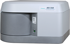
佳士科商贸有限公司为您提供《共焦拉曼成像——使用光谱学进行材料分析和化学分布》,该方案主要用于生物药品原料中材料分析、化学分布检测,参考标准--,《共焦拉曼成像——使用光谱学进行材料分析和化学分布》用到的仪器有JASCONRS5000/7000共聚焦激光拉曼光谱仪
相关方案
更多
该厂商其他方案
更多