方案详情
文
生物制药从研发到消费者可用性的道路是一个漫长而彻底的过程,旨在确保有效和质量控制。生物仿制药的开发需要与原始生物制药一起评估分子的高阶结构(HOS)。此外,生物制药的结构对制造过程或储存环境的变化很敏感,有必要评估这些分子在任何加工或储存改变后所保持的更高阶结构。
关键词:新型冠状病毒,SARS-CoV-2,新冠肺炎,生物制药,抗体药物,生物相似,VHH抗体,HOS,二级结构,三级结构,圆二色性
方案详情

CD光谱法被广泛用于评估生物制药中的HOS,因为远紫外线CD对蛋白质主链和二级结构的键角变化敏感,而近紫外线监测由芳香氨基酸残基和二硫键组成的三级结构。在这里,我们使用[光谱QC测试]程序评估了三种新型抗冠状病毒(SARS-CoV-2) VHH抗体的HOS,该程序根据CD光谱形状的差异定量确定结构变化的显著性。Application Note200-CD-036 Evaluation of HOS identity and VHH antibody binding tocoronavirus using circular dichroism spectroscopy2/5Application Note Evaluation of HOS identity and VHH antibody binding to coronavirus using Circular Dichroism Spectroscopy In t rod uc tion T h e path of a biopharmaceutical from resea r ch and development to consume r availability is a l ong and thorough process, i ntended to ensure eff i cacious and qual i ty control . Development of biosimi l ars requires eva l uation of the molecu l e's h igher order structu r e (HOS) with the or i g i nal biopharmaceutical . Additio n ally, the structure of biopharmaceuticals i s sensitive to c h anges i n the ma n u f acturing process or storage environment and it is necessary to evaluate the higher order st r ucture of these molecules sustained after any processing or storage alteratio n s. CD spect r oscopy is widely used to assess HOS i n biopharmaceuticals because t h e far-UV CD i s sensitive to changes i n the bond angles of a protein's backbone chai n and secondary st r ucture and the near-UV monitors t h e tertia r y structure, com p osed of aro m a t ic amino acids r esidues and d i sulfide bond s . Here, we evaluate the H OS o f three J-1500 CD Spec t rometer t ypes of new an t i-coronavirus (SARS-CoV-2) VHH antibodies using the [Spectral QC test] program that quantitatively determines the significance in structu r e changes based on the difference i n CD spect r al shape. K e ywor ds New corona virus, SARS-CoV-2, COVID-19, Bio p harmaceu ti cals, Antibody drug, BioSimilar, VHH antibody, HOS, Secondary Structure, Tertiary Structu r e, Ci r cular Dichroism Ex pe rim e ntal Far-UV Measurement Conditions Near-UV Measurement Conditions Wavelength 250-200nm Bandwidth 1nm Wavelength 310-250nm Bandwidth 1nm D.I.T. 4 sec Scanning Speed 20 nm/min D.I.T. 4 sec Scanning Speed 50 nm/min Data Interval 0.1nm Accumulations 1 Data Interval 0.1nm Accumulations 4 Determination of differences in CD spectra Far -UV CD meas ur eme n ts were acquired for 3 kinds of a n ti -SARS-CoV -2 VHH a ntibodies in 20 mM PBS b uf f er using the f ollowing co n cen tr atio n s: No.1(0.2 mg/mL ), No.2 (0.2 mg/mL ), No.3 (0.2 mg/mL ). Near -UV CD measu r e ments we r e o b tai n ed o n the same sa m p l es u s i n g t he following concentrations: No.1 (6.27 mg/mL ), N o .2 (7.22 mg/mL), No.3 (5.15 mg/mL ). Al l VHH ant ib ody sam pl es wer e provid e d by R e PHAGEN3. Figure 1 shows the method analysis steps for determ i ning t h e CD spect r um dif f erence between the refe r ence and t h e s a mple in the [Spectrum QC Test] pr ogram, as explained below. (1) Calculation of correlation coefficient: The CD spectru m of the refe r ence sample is measured multiple times, and the correlation coeffic i ent between t h e ave r age spectrum R (X) and the CD spectrum U () of the sam p le is calculated. (2) Weighting: The [Spectral QC Test ] program allows the user to choose whether to weight the spectrum. The CD spect r um has a low S/N due to weak li ght energy detected in t he shorter wavelength region as well as the wavelength r egio n where sample absorption is strong. Therefore, i t i s possible to reduce the we i ghting of the spectrum i n the wave l ength ra n ge where the effects f rom noise are large and increase the weighting in the wavele n gth r ange where n oise is small (Patent No. 6244492). As a result of th i s data processing, the infl u e n ce of noise can be r educed in the wave l ength ra n ge where t h e S/N i s smaller, and the spec t ral difference between samples can be determined with high sens i tivity. (3) Quantify t he difference (i .e. distance ()) between t h e r eference and sample CD spectra. (4) Judgment : T h e Z -test deter min es if there i s a sig n ificant di f f erence b e tween t h e refe r ence and sam p le spectra . I f t he Z -scor e i s 2 or less, t h e r e f e rence a n d t h e sample ar e j udg e d to be the same, and i f the Z -score is l arger t h a n 2, t h e r efere n ce and t h e sample a r e ju dged to b e d i ffere n t . Figure 1. A nalys i s s tep s for d et e r m ini ng t h e sp e ctr a l d i f f e r e n c e s bet we e n the r e f e ren ce a nd t h e s a mple s p ec tr a . Results Visual comparison of CD spectra F i gure 2 shows t h e CD spectra of VHH ant i bodies i n t h e far -UV region and n ear -UV r egio n . In t h e fa r-UV region, a n e g a tive s i g n al at 217 nm cha r act e rist i c of B-sh e et structure was observed o nl y for sample No. 2 and no significant diffe r e nce was obs e rved in t h e CD sp e ctra for samples No. 1 a n d No. 3. In the nea r-UV CD spect r a, signals d e riv e d from phenylalanine (Phe) at 255 to 270 n m , tyrosine (Ty r ) at 275 to 282 nm, and tr yptophan (Trp) at 290 to 305 n m were observed in all samples. Wh i le the CD spectra of sam p les No. 2 and No. 3 are vis u ally si mil ar, t h e spectra of sample No. 1is clearly different. Figure 2. F ar -U V (le ft) a nd n ear-U V (r igh t) CD sp e ctr a o f VH H an t ib o d i e s f or s a m p les No. 1, 2, a n d 3. Judgment of slight CD spectrum difference by statistical method T h e diff ere nc e s i n the f a r -UV CD s p ectra of sam p l e s No. 1 and No. 3 an d t he n ear -UV CD spec tr a of sampl e s No. 2 and No. 3 cannot be clearly discriminated. However, using t he [Spect r um QC T e st ] p rogram , the dista n ce b etween t h e spectra wa s obtai n ed by the a n alysis met h od shown in F i gu r e 1, and th e differences judged by the Z-test . The number of spect r a l data used for the a n alysis i s as f ollows a n d the i ndividual spect r a a r e sh o wn i n F i gure 3: F a r -UV region: Reference No. 1(9 spec tr a), sample No. 3 (5 spec t ra) Nea r-UV region : Reference No. 2 (9 spec tr a), sample No. 3 (5 spect r a) Figure 3. F ar - U V (l e f t ) a nd n ea r-U V ) ri ght C D sp e c t ra o f t he V H H ant i bod i es a nal y z e d us i n g t he [Spectr u m QC T e st ] pr o g ram. The dista n ce between the refe r ence and sample CD spectra is shown i n Figure 4 and the correspond i ng values are shown in Table 1. From Figure 4 it can be seen that weighting both the far-UV and the near-UV spectra inc r eases the distance between r eference and sample and the difference between them can be clearly confirmed. JASCO INC. 28600 Mary's C o ur t , E asto n, MD 21601US A T el : (800) 333-5272 F ax: (410) 822-7526A ppl ic a tio n Lib r ary: ja s c o i n c.com /appli c a t i o ns Table 1. Dista n ce s bet we en the r ef e ren c e a n d s ample f a r-U V (le ft ) a n d n e ar -U V (r igh t ) CD spec t r a. Far-UV Reference (No.1) Sample (No.3) Near-UV Reference (No.1) Sample (No. 3) No. ofMeasurement Weighted No weighting Weighted No weighting No. ofMeasurement Weighted No weighting Weighted No weighting 1 0.9984 0.9987 0.9916 0.9935 1 0.99980 0.99973 0.9955 0.9956 2 0.9983 0.9985 0.9902 0.9921 2 0.99988 0.99984 0.9955 0.9954 3 0.9978 0.9983 0.9929 0.9945 3 0.99986 0.99982 0.9955 0.9954 4 0.9972 0.9968 0.9914 0.9928 4 0.99986 0.99983 0.9954 0.9955 5 0.9989 0.9988 0.9920 0.9937 5 0.99987 0.99983 0.9953 0.9952 6 0.9984 0.9987 6 0.99984 0.99978 7 0.99986 0.99981 7 0.9974 0.9973 8 0.9972 0.9974 8 0.99984 0.99979 9 0.9981 0.9982 9 0.99982 0.99976 In addition, the Z -scores are shown i n Table 2. The Z -scores were all larger than 2 with and w i t h ou t weig h ting , indicat i ng tha t there wa s a significant d if fere n ce between the CD spectrum of the reference and sample both in t he far- a n d n ear-UV. Table 2. C alc ul ate d Z -s c o re s from f a r-U V a n d n ea r -U V C D s p e ct ra u s i ng [S p ect r um QC Te st ] p r o g ram. No. ofMeasurement Far-UV Near-UV Weighted No weighting Weighted No weighting 1 11.5 6.8 184 123 2 13.9 9.0 183 126 3 9.0 5.4 184 126 4 11.7 7.9 187 124 5 10.6 6.6 194 134 R efere nc e 1) W. F. W. 4th,J. P. Gabrielson, W. Al-Azzam, G. Chen, D. L. Davis, T . K. Das, D. B. Hayer, D. Houde, S. K, Technical Decision Mak i ng With Hig h er Orde r Struc t u re Data: Perspectives on Higher Orde r Structure Characterization From the Biopharmaceut i cal Industry, J. Pha r m. Sci .,105 (2016) 3465-3470 2) M. T. Gu t ier r ez Lugo, U.S. Food and Drug Administ r ation,Regulatory Consideration for the Characterizat i on of HOS in Biotechno l ogy Products, 5th I n ternationa l Sympos i um on Higher Order Structure of Protein Therapeutics 2016 3) RePHAGEN Co., Ltd (h t tps://rephagen.com/, May 20, 2020) 4) S. M. Kelly, N. C. Pric e, T h e Use of Circular Dichroism in t h e I nvestigation of Protein Structure a n d Function, Current Protein and Pept i d e Sc i ence , 1(2000) 349-384. JASCO INC. USGO A ppl ic a tio n Lib r ary: ja s c o i n c.com /appli c a t i o ns
确定


还剩3页未读,是否继续阅读?
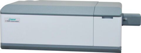
佳士科商贸有限公司为您提供《使用圆二色谱法评估HOS身份和VHH抗体与冠状病毒的结合》,该方案主要用于生物药品药物研发中分子结构检测,参考标准--,《使用圆二色谱法评估HOS身份和VHH抗体与冠状病毒的结合》用到的仪器有JASCO圆二色光谱仪CD J-1500
相关方案
更多
该厂商其他方案
更多


