


方案详情
文
Lack of TCF2vHNF1 in mice leads to
pancreas agenesis
C. Haumaitre*, E. Barbacci*, M. Jenny?, M. O. Ott*, G. Gradwohl?,and S. Cereghini*?
*Biologie du De′ veloppement, Unite′ Mixte de Recherche 7622,Centre National de la Recherche Scientifique, Universite′ Pierre etMarie Curie, 9 Quai St.
Bernard Ba t C, 75005 Paris, France; and ?Institut National de laSante′ et de la Recherche Me′ dicale U381, 3 Avenue Molie` re,67200 Strasbourg, France
Edited by Kathryn V. Anderson, Sloan–Kettering Institute, New York,NY, and approved December 20, 2004 (received for review August 6,2004)
Heterozygous mutations in the human POU-homeobox TCF2
方案详情

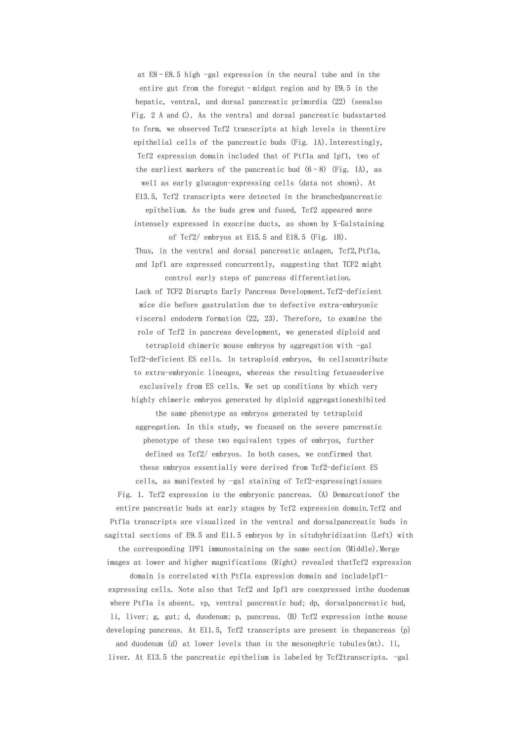
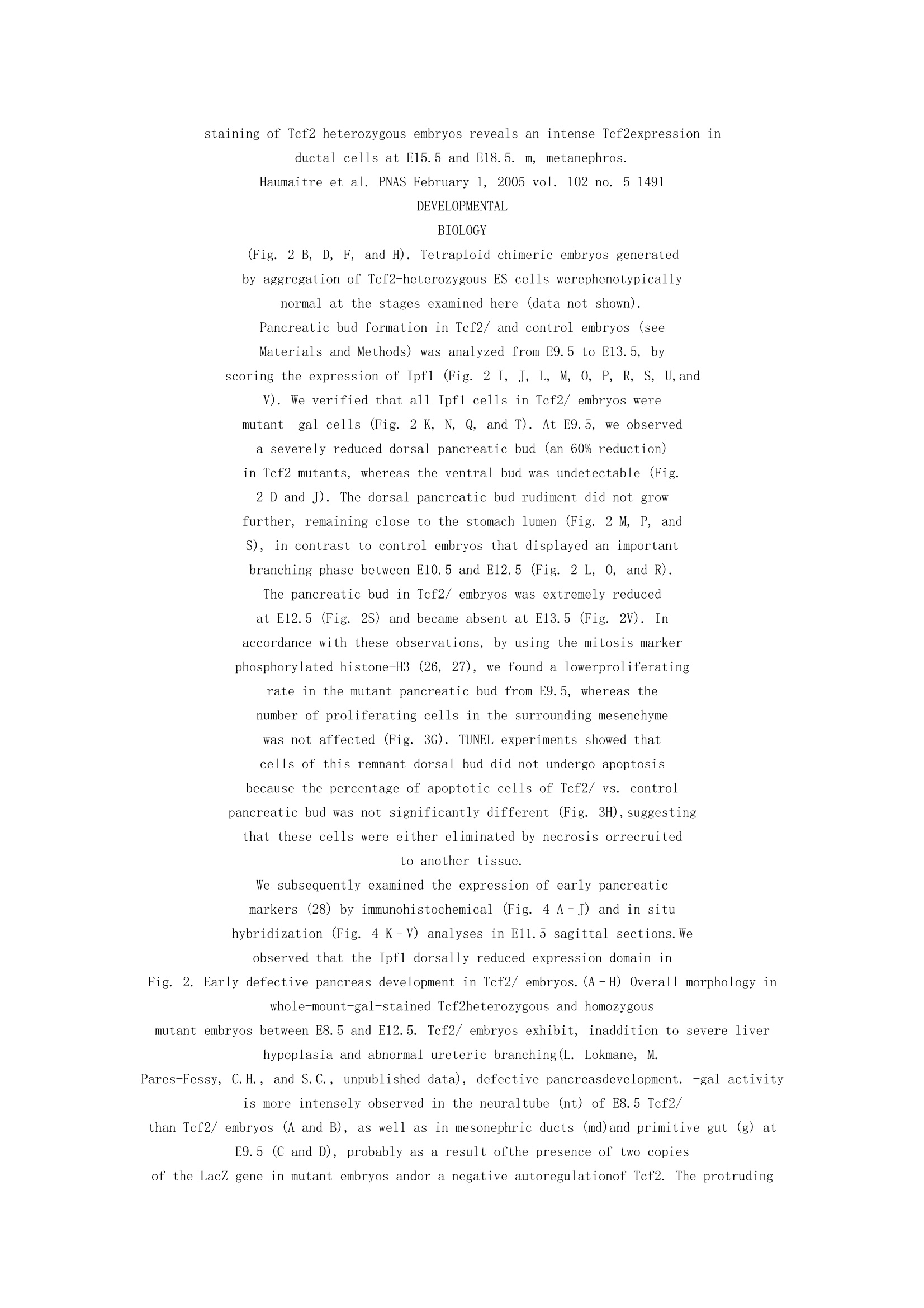
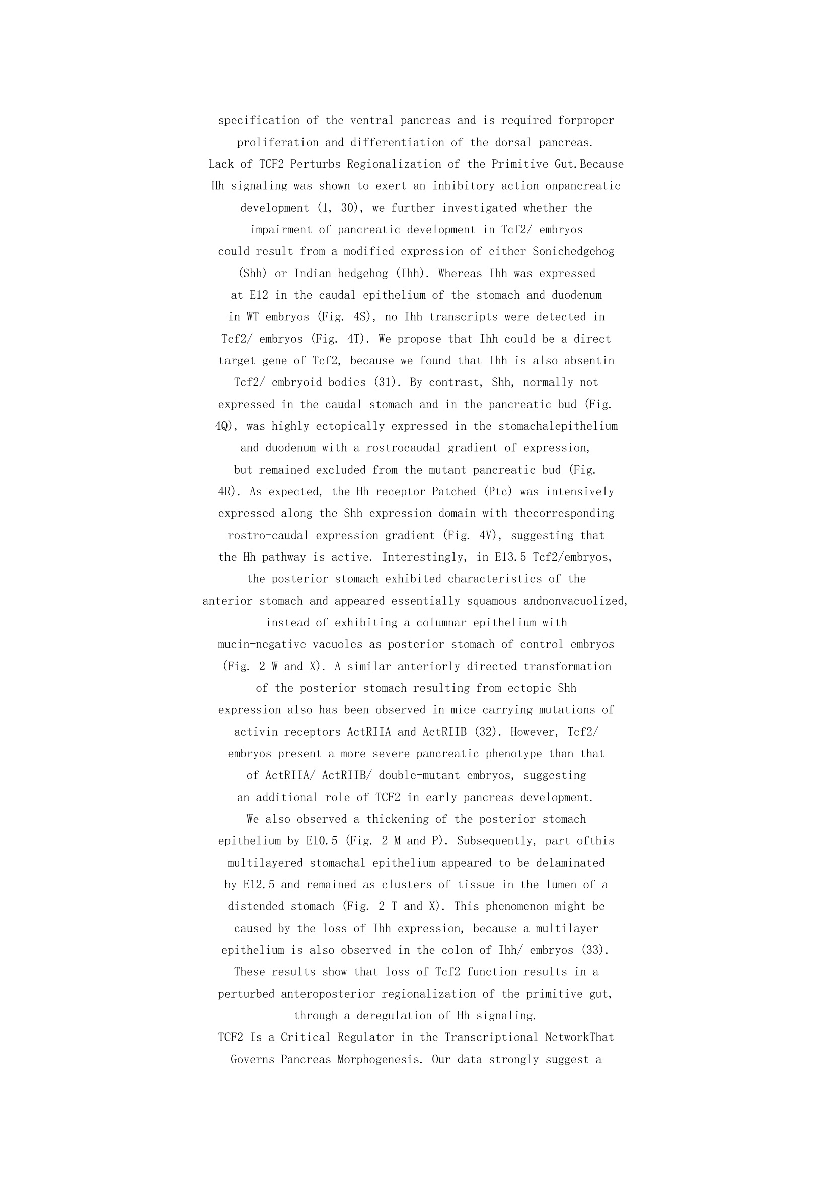
基因缺失造成鼠胰腺发育不全 Lack of TCF2vHNF1 in mice leads to pancreas agenesis C. Haumaitre*, E. Barbacci*, M. Jenny†, M. O. Ott*, G. Gradwohl†,and S. Cereghini*‡ *Biologie du De´ veloppement, Unite´ Mixte de Recherche 7622,Centre National de la Recherche Scientifique, Universite´ Pierre etMarie Curie, 9 Quai St. Bernard Ba t C, 75005 Paris, France; and †Institut National de laSante´ et de la Recherche Me´ dicale U381, 3 Avenue Molie` re,67200 Strasbourg, France Edited by Kathryn V. Anderson, Sloan–Kettering Institute, New York,NY, and approved December 20, 2004 (received for review August 6,2004) Heterozygous mutations in the human POU-homeobox TCF2 (vHNF1, HNF1) gene are associated with maturity-onset diabetes of the young, type 5, and abnormal urogenital tractdevelopment. Recently, pancreas atrophies have been reported in severalmaturity- onset diabetes of the young type 5 patients, suggesting that TCF2 is required not only for adult pancreas function but alsofor its normal development. Tcf2-deficient mice die beforegastrulation because of defective visceral endoderm formation. Toinvestigate the role of this factor in pancreas development, we rescued this early lethality by tetraploid aggregation. We show thatTCF2 has an essential function in the first steps of pancreasdevelopment, correlated with its expression domain that demarcates the entire pancreatic buds from the earliest stages. Lack of TCF2results in pancreas agenesis by embryonic day 13.5. At earlier stages,only a dorsal bud rudiment forms transiently and expresses thetranscription factors Ipf1 and Hlxb9 but lacks the key transcription factor involved in the acquisition of a pancreatic fate, Ptf1a, aswell as all endocrine precursor cells. Regional specification of thegut also is perturbed in Tcf2/ embryos as manifested by ectopic expression of Shh and lack of Ihh and Ipf1 in the posteriorstomach and duodenum. Our results highlight the requirement of Tcf2 for ensuring both accurate expression of key regulator molecules in the stomach–duodenal epithelium and proper acquisition of the pancreatic fate. This study provides further insights intoearly molecular events controlling pancreas development and maycontribute to the development of cell-replacement strategies for diabetes. diabetes MODY5 homeodomain transcription factor pancreas development gut regionalization tetraploid aggregation In mammals, the pancreas emerges as ventral and dorsal evaginations from the foregut–midgut junction that subsequently fused to form a complex organ. The signaling molecule Sonic Hedgehog (SHH) demarcates a molecular boundary between the prepancreatic endoderm and adjacent stomach and duodenal anlagen and exerts an inhibitory action on pancreas development (1–3). Genetic studies in mice have identified a hierarchical regulatory network involved in pancreasmorphogenesis, with significant and sequential differences between ventral and dorsal pancreas. In the mouse, the dorsal bud appears at embryonic day 9.5 (E9.5) concomitantly with thefirst differentiated glucagon-producing cells. The homeobox gene Ipf1(Pdx1) is expressed before and during this budding, and all pancreatic cell types derive from IPF1 progenitors (4, 5). However, in Ipf1-deficient mice, pancreas development isarrested after budding (6, 7), implying that other factors promote pancreas specification. Recently, the transcription factorPtf1a (P48) has been shown to be essential for the acquisition of a pancreatic fate by undifferentiated ventral foregut endoderm, being required for the specification of the ventral pancreasand robust outgrowth of the dorsal bud. In its absence, ventral pancreas progenitors differentiate into duodenal cells bydefault (8). By contrast, the homeobox gene Hlxb9 is required only dorsally, for specifying the gut epithelium to a pancreatic fate(9, 10). A key regulator of endocrine development is the basic helix–loop–helix protein Neurogenin3 (Ngn3), which isabsolutely required to promote islet cell development (11). The Isl1 gene, which encodes a LIM-homeodomain protein, performs two functions in the developing pancreas. It is initiallyrequired in the dorsal mesenchyme for proper exocrine differentiation and later in the pancreatic epithelium for islet survival (12). Downstream of them, other transcription factors are essentialfor proper pancreatic endocrine differentiation such as Nkx2.2 and Pax6 (13–15). However, the initial stages of pancreaticdevelopment occur early in mammalian embryogenesis, and molecular mechanisms governing these first steps remain to be elucidated. In humans, mutations in the POU-homeobox TCF2 gene are associated with the human disease maturity-onset diabetes ofthe young type 5, a form of dominantly inherited type II diabetes mellitus characterized by pancreatic beta cell dysfunction atthe age of 25 years or younger, nondiabetic early onset renaldisease, liver dysfunction, and abnormal urogenital tract development (16–18). In addition to these phenotypes, variable levels of pancreas atrophies have recently been associated with different TCF2 mutations (19, 20). Remarkably, we have recentlyidentified a severe pancreas hypoplasia in two fetuses carrying previously undescribed mutations in the TCF2 gene (A. L. Delezoide, C.H., and S.C., unpublished results). These data, together with the observation that vHnf1(Tcf2)-mutant embryos show underdevelopment of the pancreas in zebrafish (21), strongly suggest a critical function of Tcf2 in pancreasdevelopment. However, the molecular bases of these pancreatic phenotypes are poorly understood, as are the Tcf2 target genes involved. In mice, the precise implication of Tcf2 during early organogenesis remains essentially unknown, because Tcf2- deficient embryos die before gastrulation due to defective visceral endoderm formation (22, 23). In this study, we rescued this early lethality by tetraploid aggregation, by using Tcf2/ embryonic stem (ES) cells. We observed in these rescued Tcf2-null embryos an absence of the ventral pancreatic bud and an extremely reduced and transient dorsal bud that leads to pancreas agenesis by E13.5. Ourresults uncover the requirement of Tcf2 for the specification of the ventral pancreas and for proper morphogenesis anddifferentiation of the dorsal pancreas. They further suggest that Tcf2 also is required for both accurate regionalization of the primitivegut through Hedgehog (Hh) signaling and proper acquisition of the pancreatic fate by regulating Ptf1a expression, thus placingthis transcription factor at one of the highest positions in thegenetic network that controls pancreas development. Materials and Methods Diploid and Tetraploid Chimera. Because chimeric embryosgenerated with our previously isolated Tcf2/ and Tcf2/ ES cells This paper was submitted directly (Track II) to the PNASoffice. Abbreviations: En, embryonic day n; ES, embryonic stem; Hh,hedgehog; vHnf1, variant Hepatocyte nuclear factor 1; Shh, Sonic Hedgehog; Ihh, IndianHedgehog; Ngn3, Neurogenin3; Isl1, Islet-1; Hnf6, Hepatocyte nuclear factor 6. ‡To whom correspondence should be addressed. E-mail: silvia.cereghini@snv.jussieu.fr (mailto:silvia.cereghini@snv.jussieu.fr). © 2005 by The National Academy of Sciences of the USA 1490–1495 PNAS February 1, 2005 vol. 102 no. 5 www.pnas.orgcgidoi10.1073pnas.0405776102 (http://www.pnas.org%01cgi%01doi%0110.1073%01pnas.0405776102/) presented neural tube defects that were inherent to theparental cell line (22), we isolated seven previously undescribed EScell lines (four Tcf2/ and three Tcf2/) from blastocysts obtained after crossing Tcf2-heterozygous mice (129sv background), as described in ref. 24. Tetraploid embryos were generated by electrofusion on a cell fusion instrument (CF-150, BLS Ltd., Budapest; voltage, 80 V; duration, 80 ms; 1 pulse) at thetwo-cell stage. Tetraploid or diploid chimeric embryos were generated as described in ref. 25. Two four-cell stage CD1 tetraploidembryos or a wild-type (WT) CD1 morula were aggregated with a single loose clump of 15–20 Tcf2-deficient ES cells, cultured in M16 medium (Sigma) up to the blastocyst stage, and implanted into pseudopregnant females. We first confirmed that tetraploid or diploid embryos generated with our Tcf2/ ES cells were similar to WT or heterozygous Tcf2 embryos, indicating that thephenotype of Tcf2/ ES cells-derived embryos is specifically due to the lack of TCF2. We used as control embryos in a given litter blastocysts obtained from cultured morulae not aggregated with ES cells and implanted together with ES-cell-aggregatedembryos. Because the yield of tetraploid embryos was low, we established optimal conditions to obtain diploid chimera with maximal ES-cell contribution, and we verified that these very highly diploid chimeric embryos displayed the same phenotype as tetraploid embryos. Because we disrupted the Tcf2 gene by inserting the LacZ gene, the relative contribution of WT and mutant cells in ES-cell-derived embryos was evaluated bywholemount X-Gal staining (22). We analyzed here only very highly chimeric and tetraploid embryos, characterized by the presence of exclusively -gal mutant cells in the Tcf2-expressing tissues (defined as Tcf2/ embryos). Immunohistochemistry, in Situ Hybridization, and TUNEL. Mouse embryos were fixed in 4% paraformaldehyde and embedded in paraffin. Then, 5-msagittal sections were dewaxed, rehydrated, and subjected to microwave antigen retrieval in 10 mM citrate. For immunostaining, we used rabbit anti-Ipf1 (M. German, Hormone Research Institute, San Francisco), mouse antiglucagon (Sigma), rabbit anti-Hlxb9 (10), mouse anti-Islet-1 (39.4D5 and 40.2D6), mouse anti-Pax6 (Developmental Studies Hybridoma Bank, Iowa City, IA), and rabbit anti-phosphohistone H3 (Upstate Biotechnology, Lake Placid, NY) as primary antibodies, and FITC- and Cyanine3-conjugated (The Jackson Laboratory) as secondary antibodies. For in situ hybridization, we prepared frozen sections from timed embryos, as described in ref. 11. The following cRNA probes were used: Ptf1a (P. Wellauer, Swiss Institute for Experimental Cancer Research, Epalinges, Switzerland); Hnf6 (F. Lemaigre, Universite´ Catholique de Louvain, Brussels); Ngn3 (11); Shh and Ihh (A. P. McMahon, Harvard University, Cambridge, MA); and Ptc (M. Scott, Howard Hughes Medical Institute, Stanford, CA). TUNEL was performed by using the fluorescein cell death detection kit (Roche). Results and Discussion Tcf2 Is Expressed in the Developing Pancreas from Its EarlyStages. Tcf2 heterozygous embryos for a null allele with the LacZ gene under the control of regulatory regions of the Tcf2 locusexhibit at E8–E8.5 high -gal expression in the neural tube and in the entire gut from the foregut–midgut region and by E9.5 in the hepatic, ventral, and dorsal pancreatic primordia (22) (seealso Fig. 2 A and C). As the ventral and dorsal pancreatic budsstarted to form, we observed Tcf2 transcripts at high levels in theentire epithelial cells of the pancreatic buds (Fig. 1A).Interestingly, Tcf2 expression domain included that of Ptf1a and Ipf1, two of the earliest markers of the pancreatic bud (6–8) (Fig. 1A), as well as early glucagon-expressing cells (data not shown). At E13.5, Tcf2 transcripts were detected in the branchedpancreatic epithelium. As the buds grew and fused, Tcf2 appeared more intensely expressed in exocrine ducts, as shown by X-Galstaining of Tcf2/ embryos at E15.5 and E18.5 (Fig. 1B). Thus, in the ventral and dorsal pancreatic anlagen, Tcf2,Ptf1a, and Ipf1 are expressed concurrently, suggesting that TCF2 might control early steps of pancreas differentiation. Lack of TCF2 Disrupts Early Pancreas Development.Tcf2-deficient mice die before gastrulation due to defective extra-embryonic visceral endoderm formation (22, 23). Therefore, to examine the role of Tcf2 in pancreas development, we generated diploid and tetraploid chimeric mouse embryos by aggregation with -gal Tcf2-deficient ES cells. In tetraploid embryos, 4n cellscontribute to extra-embryonic lineages, whereas the resulting fetusesderive exclusively from ES cells. We set up conditions by which very highly chimeric embryos generated by diploid aggregationexhibited the same phenotype as embryos generated by tetraploid aggregation. In this study, we focused on the severe pancreatic phenotype of these two equivalent types of embryos, further defined as Tcf2/ embryos. In both cases, we confirmed that these embryos essentially were derived from Tcf2-deficient ES cells, as manifested by -gal staining of Tcf2-expressingtissues Fig. 1. Tcf2 expression in the embryonic pancreas. (A) Demarcationof the entire pancreatic buds at early stages by Tcf2 expression domain.Tcf2 and Ptf1a transcripts are visualized in the ventral and dorsalpancreatic buds in sagittal sections of E9.5 and E11.5 embryos by in situhybridization (Left) with the corresponding IPF1 immunostaining on the same section (Middle).Merge images at lower and higher magnifications (Right) revealed thatTcf2 expression domain is correlated with Ptf1a expression domain and includeIpf1- expressing cells. Note also that Tcf2 and Ipf1 are coexpressed inthe duodenum where Ptf1a is absent. vp, ventral pancreatic bud; dp, dorsalpancreatic bud, li, liver; g, gut; d, duodenum; p, pancreas. (B) Tcf2 expression inthe mouse developing pancreas. At E11.5, Tcf2 transcripts are present in thepancreas (p) and duodenum (d) at lower levels than in the mesonephric tubules(mt). li, liver. At E13.5 the pancreatic epithelium is labeled by Tcf2transcripts. -gal staining of Tcf2 heterozygous embryos reveals an intense Tcf2expression in ductal cells at E15.5 and E18.5. m, metanephros. Haumaitre et al. PNAS February 1, 2005 vol. 102 no. 5 1491 DEVELOPMENTAL BIOLOGY (Fig. 2 B, D, F, and H). Tetraploid chimeric embryos generated by aggregation of Tcf2-heterozygous ES cells werephenotypically normal at the stages examined here (data not shown). Pancreatic bud formation in Tcf2/ and control embryos (see Materials and Methods) was analyzed from E9.5 to E13.5, by scoring the expression of Ipf1 (Fig. 2 I, J, L, M, O, P, R, S, U,and V). We verified that all Ipf1 cells in Tcf2/ embryos were mutant -gal cells (Fig. 2 K, N, Q, and T). At E9.5, we observed a severely reduced dorsal pancreatic bud (an 60% reduction) in Tcf2 mutants, whereas the ventral bud was undetectable (Fig. 2 D and J). The dorsal pancreatic bud rudiment did not grow further, remaining close to the stomach lumen (Fig. 2 M, P, and S), in contrast to control embryos that displayed an important branching phase between E10.5 and E12.5 (Fig. 2 L, O, and R). The pancreatic bud in Tcf2/ embryos was extremely reduced at E12.5 (Fig. 2S) and became absent at E13.5 (Fig. 2V). In accordance with these observations, by using the mitosis marker phosphorylated histone-H3 (26, 27), we found a lowerproliferating rate in the mutant pancreatic bud from E9.5, whereas the number of proliferating cells in the surrounding mesenchyme was not affected (Fig. 3G). TUNEL experiments showed that cells of this remnant dorsal bud did not undergo apoptosis because the percentage of apoptotic cells of Tcf2/ vs. control pancreatic bud was not significantly different (Fig. 3H),suggesting that these cells were either eliminated by necrosis orrecruited to another tissue. We subsequently examined the expression of early pancreatic markers (28) by immunohistochemical (Fig. 4 A–J) and in situ hybridization (Fig. 4 K–V) analyses in E11.5 sagittal sections.We observed that the Ipf1 dorsally reduced expression domain in Fig. 2. Early defective pancreas development in Tcf2/ embryos.(A–H) Overall morphology in whole-mount-gal-stained Tcf2heterozygous and homozygous mutant embryos between E8.5 and E12.5. Tcf2/ embryos exhibit, inaddition to severe liver hypoplasia and abnormal ureteric branching(L. Lokmane, M. Pares-Fessy, C.H., and S.C., unpublished data), defective pancreasdevelopment. -gal activity is more intensely observed in the neuraltube (nt) of E8.5 Tcf2/ than Tcf2/ embryos (A and B), as well as in mesonephric ducts (md)and primitive gut (g) at E9.5 (C and D), probably as a result ofthe presence of two copies of the LacZ gene in mutant embryos andor a negative autoregulationof Tcf2. The protruding dorsal pancreatic bud externally detectedin heterozygous embryos between E9.5 and E12.5 is not observed in homozygous mutantembryos (black arrowhead, C–H). (I–X) Pancreatic bud morphogenesisin sagittal sections of control and Tcf2 homozygous mutant embryos between E9.5 andE13.5. (I–K) IPF1 immunostainings of sagittal sections ofwhole-mount-gal-stained embryos reveal ventral and dorsal pancreatic buds in Tcf2/ embryos, whereasthe ventral pancreatic bud is totally absent and only a veryreduced dorsal bud is observed in Tcf2/ embryos at E9.5. vp, ventral pancreas; dp, dorsalpancreas. (L–T) At later stages, the dorsal pancreatic bud (whitearrowhead), the duodenum (d), and the posterior stomach are stained by IPF1 in control embryos, butonly a remnant pancreatic bud is stained in Tcf2-deficient embryos,which is abnormally close to the posterior stomachal epithelium. Whereas the pancreatic budexhibits an important growth in control embryos particularly fromE12.5, the remnant pancreatic bud in Tcf2/ embryos regresses by E12.5 and is notfurther detected at E13.5 (pancreas agenesis) (U–X). (K, N,Q, andT) -gal staining of the remnant pancreatic bud in sagittal sections of Tcf2 homozygous mutantembryos. -gal and IPF1-stained sections are counterstained bysafranin. -gal mutant cells are detected in the rudiment of the pancreatic bud in Tcf2/ embryoscoexpressing IPF1 and display a broader expression domain includinga thickness of the stomachal epithelium between E10.5 and E11.5. Note that themagnification in K, N, Q, and T is higher than in the correspondingIPF1-stained section in J, M, P, and S. (U–X) Stomachal epithelium morphology in E13.5 Tcf2control and homozygous mutant embryos. Trichromic staining of theIPF1-stained sagittal sections. Arrows indicate the posterior stomachal epithelium, whichis surrounded by the stomachal mesenchyme. Whereas the normalposterior stomach exhibits a columnar vacuolized epithelium, the posterior stomach of Tcf2/embryos appears squamous and nonvacuolized, as is normally theanterior stomach. In M, P, T, and X, an asterisk indicates thickening of the gastricepithelium. 1492 www.pnas.orgcgidoi10.1073pnas.0405776102 (http://www.pnas.org%01cgi%01doi%0110.1073%01pnas.0405776102/)Haumaitre et al. mutant embryos also expressed the early pancreatic marker Hlxb9 (Fig. 4 B and D). By contrast, both Ipf1 and Hlxb9 expression were not detected in the presumptive ventralpancreatic bud area. Remarkably, we found no expression of the key transcription factor Ptf1a (8) (Fig. 4L). Moreover, Tcf2/ mutants displayed a very reduced expression of Hnf6 and no Ngn3 expression in the remnant dorsal pancreatic bud, two factors required for endocrine fate acquisition (11, 29) (Fig. 4N and P). Consistent with this absence of Ngn3, the earliestknown marker of endocrine precursors (11), expressions of Isl1, Pax6, and glucagon were lost (Fig. 4 F, H, and J). Isl1 remained, however, expressed in the mesenchyme (7) (Fig. 4F), a tissue where Tcf2 was not expressed. Thus, endocrine precursors are totally absent in Tcf2/ pancreatic epithelium. Taken together, our results show that Tcf2 controls initial specification of the ventral pancreas and is required forproper proliferation and differentiation of the dorsal pancreas. Lack of TCF2 Perturbs Regionalization of the Primitive Gut.Because Hh signaling was shown to exert an inhibitory action onpancreatic development (1, 30), we further investigated whether the impairment of pancreatic development in Tcf2/ embryos could result from a modified expression of either Sonichedgehog (Shh) or Indian hedgehog (Ihh). Whereas Ihh was expressed at E12 in the caudal epithelium of the stomach and duodenum in WT embryos (Fig. 4S), no Ihh transcripts were detected in Tcf2/ embryos (Fig. 4T). We propose that Ihh could be a direct target gene of Tcf2, because we found that Ihh is also absentin Tcf2/ embryoid bodies (31). By contrast, Shh, normally not expressed in the caudal stomach and in the pancreatic bud (Fig. 4Q), was highly ectopically expressed in the stomachalepithelium and duodenum with a rostrocaudal gradient of expression, but remained excluded from the mutant pancreatic bud (Fig. 4R). As expected, the Hh receptor Patched (Ptc) was intensively expressed along the Shh expression domain with thecorresponding rostro-caudal expression gradient (Fig. 4V), suggesting that the Hh pathway is active. Interestingly, in E13.5 Tcf2/embryos, the posterior stomach exhibited characteristics of the anterior stomach and appeared essentially squamous andnonvacuolized, instead of exhibiting a columnar epithelium with mucin-negative vacuoles as posterior stomach of control embryos (Fig. 2 W and X). A similar anteriorly directed transformation of the posterior stomach resulting from ectopic Shh expression also has been observed in mice carrying mutations of activin receptors ActRIIA and ActRIIB (32). However, Tcf2/ embryos present a more severe pancreatic phenotype than that of ActRIIA/ ActRIIB/ double-mutant embryos, suggesting an additional role of TCF2 in early pancreas development. We also observed a thickening of the posterior stomach epithelium by E10.5 (Fig. 2 M and P). Subsequently, part ofthis multilayered stomachal epithelium appeared to be delaminated by E12.5 and remained as clusters of tissue in the lumen of a distended stomach (Fig. 2 T and X). This phenomenon might be caused by the loss of Ihh expression, because a multilayer epithelium is also observed in the colon of Ihh/ embryos (33). These results show that loss of Tcf2 function results in a perturbed anteroposterior regionalization of the primitive gut, through a deregulation of Hh signaling. TCF2 Is a Critical Regulator in the Transcriptional NetworkThat Governs Pancreas Morphogenesis. Our data strongly suggest a critical role for Tcf2 in the orchestratred network oftranscription factors and secretory molecules (34) controlling the expansionof endodermal progenitors and their differentiation intopancreatic primordia. Remarkably, Tcf2-deficient embryos exhibit a very close phenotype to that caused by Ptf1a deficiency, in regard to the absence of a ventral pancreas and a reduced dorsal pancreas. Intriguingly, we identified a TCF12-DNA consensus-binding site (ATTAATGTTTAAC) in the Ptf1a promoter at 5,092 bp from the initiation site, within a domain highly conserved between mouse and human, which specifically binds TCF12 proteins (data not shown). This finding suggests that TCF2 could regulate Ptf1a expression directly. However, thephenotype of Tcf2/ embryos is more severe than that of Ptf1a/ embryos, because in the absence of Ptf1a expression the dorsal pancreas is maintained and relatively developed, with endocrine cells still present (8, 35), indicating that other regulatory factors may contribute to Tcf2 mutant phenotype. In this context, Ipf1 and Ngn3 were previously identified as direct target genes of TCF2 (36, 37). Yet, Ipf1 was still detectablein the Tcf2/ pancreatic bud rudiment (Fig. 2), implying that TCF2 is not required for Ipf1 initial induction. By contrast,the expression of Ngn3, a gene whose expression was transiently reduced in Hnf6/ embryos (29, 38), was completely abolished in Tcf2/ embryos. Because Hnf6 expression also was Fig. 3. Decreased cell proliferation in pancreas of Tcf2/ embryos.(A and B) The pancreatic bud in control and Tcf2-homozygous mutant embryos isdefined by Ipf1 expression domain. (C and D) The mitosis markerantiphosphorylated histone H3 (P-H3) antibody detects proliferating cells in thepancreatic bud (circled by white dashed lines). (E and F) TUNEL experimentsshow cells in apoptosis in control and Tcf2-mutant pancreas (circled by whitedashed lines). (G) The percentage of P-H3 cells among IPF1 cells of control andTcf2/ pancreatic buds reveals an important cell proliferation decrease inTcf2- mutant pancreas. (H) The percentage of TUNEL cells among IPF1cells reveals no significant difference between control and Tcf2/pancreatic buds. A total of 13 sections from control (n 5) and 7 sections from Tcf2/(n 4) of E9.5 and E10.5 embryos were evaluated. Haumaitre et al. PNAS February 1, 2005 vol. 102 no. 5 1493 DEVELOPMENTAL BIOLOGY severely reduced in Tcf2/ dorsal pancreatic bud, activation of Ngn3 may require the concurrent action of Tcf2 and Hnf6. Recent studies have reported a transient reduction in TCF2 levels in the pancreatic duct cells in Hnf6-deficient embryos from E13.5 to E15.5, leading to the suggestion that Hnf6 is upstream of Tcf2 (38). Nevertheless, the more severe pancreatic phenotype of Tcf2-mutant embryos, compared with that of Hnf6/ embryos (29), indicates that this transcriptional hi- Fig. 5. Proposed model for Tcf2 function in the development of thepancreas and gut endoderm. (A) Expression domains of thetranscription factors Tcf2, Ipf1, and Ptf1a, as well as the signaling molecules Shh and Ihh, in thefore-midgut area of Tcf2/ and Tcf2/ embryos. Tcf2 deficiency leadsto an extremely reduced pancreatic bud expressing Ipf1 but missing Ptf1a expression,associated with a perturbed gut regionalization reflected by anexpansion of Shh expression domain and an absence of Ihh and Ipf1 expression in stomach and duodenum.,expressed gene;, nonexpressed gene. (B) Proposed model of theregulatory network that governs differentiation of pancreatic cells. The diagramillustrates the epistatic relations of genes required for pancreasdifferentiation, leading to endocrine and exocrine cells. Tcf2 is required early in pancreas development,activating Ptf1a, and regulating the Ipf1-Shh network, with Shhrepression vs. Ihh activation. TCF2 also activates Ngn3 in endocrine precursor cells, in agreementwith the hypothesis that TCF2-positive cells are the precursors ofNGN3 positive cells (38). Fig. 4. Impaired early pancreatic cell differentiation in Tcf2/embryos. (A–J) Immunohistochemical analysis of E11.5 control andTcf2/ dorsal pancreatic buds. Anti-Ipf1 (A and B), anti-Hlxb9 (C and D), anti-Isl1 (E andF), anti-Pax6 (G and H), and anti-glucagon (I and J) antibodieswere used on sagittal sections of WT and Tcf2-mutant embryos. (A and B) Ipf1 was detected in theremnant dorsal pancreatic bud (marked by dashed lines), but not inthe duodenum (circled by a white line), of Tcf2/ embryos. (C and D) Hlxb9 also is expressedin this bud. (E–J) None of the early endocrine pancreatic markers,Isl1, Pax6, and glucagon, were detected in the dorsal pancreatic epithelium of Tcf2 mutants.(E and F) Isl1, although absent in pancreatic epithelial cellclusters, is still expressed in the mesenchyme. (K–V) In situ hybridization analysis of control andTcf2/ E11.5 dorsal pancreatic buds. In contrast to Ipf1, Ptf1a andNgn3 are not expressed in the remnant dorsal pancreatic bud of Tcf2 mutants (K, L, O, and P),whereas Hnf6 is severely reduced (M and N). Whereas Ihh expressionis abolished in Tcf2/ mutants (S and T), Shh is highly expressed in the anterior stomachwith an expanded expression domain in posterior stomach andduodenum (Q and R) and induced Ptc expression (U and V). 1494 www.pnas.orgcgidoi10.1073pnas.0405776102 (http://www.pnas.org%01cgi%01doi%0110.1073%01pnas.0405776102/)Haumaitre et al. erarchy most likely is not involved at earlier stages ofpancreas development. Regulatory circuits also are involved in regional specification of the gut endoderm. One important aspect of thisregionalization is the restricted expression of Ipf1 and Shh, essential to permit pancreas development (12). Several studies havesuggested that Shh and Ipf1 mutually repress their expression through a regulatory loop in the gut endoderm (1, 21, 30). In correlation with this finding, we found in Tcf2/ embryos an expanded domain of Shh expression in the posterior stomach and duodenum, whereas the Ipf1 expression domain remainedrestricted to the rudimentary dorsal pancreatic bud but was absent in posterior stomach and duodenum (Fig. 4). Thus, Tcf2 appears to regulate regional specification of the gut endoderm through the Shh-Ipf1 network. Taken together, these findings allow us to propose a model highlighting the critical role played by Tcf2 in the control of pancreas development, in relation with the regionalization ofthe primitive gut (Fig. 5). We propose that in the absence of TCF2, Ptf1a expression is not induced, leading to defectivespecification of the ventral pancreas and a reduced dorsal pancreas, which is subsequently not maintained because of an alteredregionalization of the gut through deregulation of Hh signaling. This study provides further insight into the early molecular events controlling pancreas development in mice and thefunction of the transcription factor TCF2 in this process. Our observation of pancreas hypoplasia in two fetuses carryingnovel TCF2 mutations (A. L. Delezoide, C.H, and S.C, unpublished data) suggests that decreased levels of TCF2 also perturbnormal pancreas growth and function in humans. The role played by Tcf2 in pancreas development thus appears to be conserved during evolution. Then, understanding how Tcf2 together with other regulatory molecules direct early pancreas development in mice may help to elaborate cell-replacement strategies for diabetes mellitus. We thank B. Thorens (Institute of Physiology of Lausanne,Lausanne, Switzerland), M. German, P. Wellauer, F. Lemaigre, A. P. McMahon,M. Scott, and S. Schneider-Maunoury (Unite´ Mixte de Recherche7622, Centre National de la Recherche Scientifique, Universite´ Pierre etMarie Curie) for reagents, and J. F. Colas, J. L. Duband, and S.Schneider- Maunoury for comments on the manuscript. This work was supportedby Association pour la Recherche sur le Cancer Contracts 58243231, Institut National de la Sante´ et de la Recherche Me´dicale,Centre National de la Recherche Scientifique, and Universite´ Pierre etMarie Curie. C.H. is a recipient of Ph.D. student fellowships fromMiniste`re de la Recherche et de la Technologie and Association pour la Recherchesur le Cancer. 1. Apelqvist, A., Ahlgren, U. & Edlund, H. (1997) Curr. Biol.7, 801–804. 2. Hebrok, M., Kim, S. K. & Melton, D. A. (1998) Genes Dev. 12,1705– 1713. 3. Kim, S. K., Hebrok, M. & Melton, D. A. (1997) Development(Cambridge, U.K.) 124, 4243–4252. 4. Ohlsson, H., Karlsson, K. & Edlund, T. (1993) EMBO J. 12,4251–4259. 5. Gu, G., Dubauskaite, J.&Melton, D. A. (2002) Development(Cambridge, U.K.) 129, 2447–2457. 6. Offield, M. F., Jetton, T. L., Labosky, P. A., Ray, M., Stein,R. W., Magnuson, M. A., Hogan, B. L.&Wright, C. V. (1996) Development(Cambridge, U.K.) 122, 983–995. 7. Ahlgren, U., Jonsson, J. & Edlund, H. (1996) Development(Cambridge, U.K.) 122, 1409–1416. 8. Kawaguchi, Y., Cooper, B., Gannon, M., Ray, M., MacDonald, R. J.& Wright, C. V. (2002) Nat. Genet. 32, 128–134. 9. Li, H., Arber, S., Jessell, T. M. & Edlund, H. (1999) Nat.Genet. 23, 67–70. 10. Harrison, K. A., Thaler, J., Pfaff, S. L., Gu, H. & Kehrl,J. H. (1999) Nat. Genet. 23, 71–75. 11. Gradwohl, G., Dierich, A., LeMeur, M. & Guillemot, F.(2000) Proc. Natl. Acad. Sci. USA 97, 1607–1611. 12. Ahlgren, U., Pfaff, S. L., Jessell, T. M., Edlund, T. &Edlund, H. (1997) Nature 385, 257–260. 13. Sussel, L., Kalamaras, J., Hartigan-O’Connor, D. J., Meneses,J. J., Pedersen, R. A., Rubenstein, J. L. & German, M. S. (1998) Development(Cambridge, U.K.) 125, 2213–2221. 14. St-Onge, L., Sosa-Pineda, B., Chowdhury, K., Mansouri, A. &Gruss, P. (1997) Nature 387, 406–409. 15. Sander, M., Neubuser, A., Kalamaras, J., Ee, H. C., Martin, G.R. & German, M. S. (1997) Genes Dev. 11, 1662–1673. 16. Nishigori, H., Yamada, S., Kohama, T., Tomura, H., Sho, K.,Horikawa, Y., Bell, G. I., Takeuchi, T. & Takeda, J. (1998) Diabetes 47,1354–1355. 17. Lindner, T. H., Njolstad, P. R., Horikawa, Y., Bostad, L.,Bell, G. I. & Sovik, O. (1999) Hum. Mol. Genet. 8, 2001–2008. 18. Bingham, C., Ellard, S., Allen, L., Bulman, M., Shepherd, M.,Frayling, T., Berry, P. J., Clark, P. M., Lindner, T., Bell, G. I., et al. (2000)Kidney Int. 57, 898–907. 19. Bellanne-Chantelot, C., Chauveau, D., Gautier, J.,Dubois-Laforgue, D., Clauin, S., Beaufils, S., Wilhelm, J. M., Boitard, C., Noel, L. H.,Velho, G. & Timsit, J. (2004) Ann. Intern. Med. 140, 510–517. 20. Barbacci, E., Chalkiadaki, A., Masdeu, C., Haumaitre, C.,Lokmane, L., Loirat, C., Cloarec, S., Talianidis, I., Bellanne-Chantelot, C. &Cereghini, S. (2004) Hum. Mol. Genet. 13, 3139–3149. 21. Sun, Z. & Hopkins, N. (2001) Genes Dev. 15, 3217–3229. 22. Barbacci, E., Reber, M., Ott, M., Breillat, C., Huetz, F. &Cereghini, S. (1999) Development (Cambridge, U.K.) 126, 4795–4805. 23. Coffinier, C., Thepot, D., Babinet, C., Yaniv, M. & Barra,J. (1999) Development (Cambridge, U.K.) 126, 4785–4794. 24. Hogan, B., Beddington, R. S., Costantini, F. & Lacy, E.(1994) Manipulating the Mouse Embryo: A Laboratory Manual (Cold Spring Harbor Lab. Press,Woodbury, NY). 25. Nagy, A., Rossant, J., Nagy, R., Abramow-Newerly, W. &Roder, J. C. (1993) Proc. Natl. Acad. Sci. USA 90, 8424–8428. 26. Schmiesing, J. A., Gregson, H. C., Zhou, S. & Yokomori, K.(2000) Mol. Cell. Biol. 20, 6996–7006. 27. Bort, R., Martinez-Barbera, J. P., Beddington, R. S. &Zaret, K. S. (2004) Development (Cambridge, U.K.) 131, 797–806. 28. Wilson, M. E., Scheel, D. & German, M. S. (2003) Mech. Dev.120, 65–80. 29. Jacquemin, P., Durviaux, S. M., Jensen, J., Godfraind, C.,Gradwohl, G., Guillemot, F., Madsen, O. D., Carmeliet, P., Dewerchin, M., Collen,D., et al. (2000) Mol. Cell. Biol. 20, 4445–4454. 30. Hebrok, M., Kim, S. K., St Jacques, B., McMahon, A.P.&Melton, D. A. (2000) Development (Cambridge, U.K.) 127, 4905–4913. 31. Haumaitre, C., Reber, M. & Cereghini, S. (2003) J. Biol.Chem. 278, 40933– 40942. 32. Kim, S. K., Hebrok, M., Li, E., Oh, S. P., Schrewe, H., Harmon,E. B., Lee, J. S. & Melton, D. A. (2000) Genes Dev. 14, 1866–1871. 33. Van den Brink, G. R., Bleuming, S. A., Hardwick, J. C.,Schepman, B. L., Offerhaus, G. J., Keller, J. J., Nielsen, C., Gaffield, W., vanDeventer, S. J., Roberts, D. J. & Peppelenbosch, M. P. (2004) Nat. Genet. 36,277–282. 34. Kumar, M. & Melton, D. (2003) Curr. Opin. Genet. Dev. 13,401–407. 35. Krapp, A., Knofler, M., Ledermann, B., Burki, K., Berney, C.,Zoerkler, N., Hagenbuchle, O. & Wellauer, P. K. (1998) Genes Dev. 12,3752–3763. 36. Gerrish, K., Cissell, M. A. & Stein, R. (2001) J. Biol.Chem. 276, 47775–47784. 37. Lee, J. C., Smith, S. B., Watada, H., Lin, J., Scheel, D.,Wang, J., Mirmira, R. G. & German, M. S. (2001) Diabetes 50, 928–936. 38. Maestro, M. A., Boj, S., Luco, R. F., Pierreux, C. E., Cabedo,J., Servitja, J. M., German, M. S., Rousseau, G. G., Lemaigre, F. P.&Ferrer, J.(2003) Hum. Mol. Genet. 12, 3307–3314. Haumaitre et al. PNAS February 1, 2005 vol. 102 no. 5 1495 DEVELOPMENTAL BIOLOGY
确定


还剩11页未读,是否继续阅读?

产品配置单
香港友诚生物科技有限公司为您提供《基因缺失造成鼠胰腺发育不全》,该方案主要用于其他中--检测,参考标准--,《基因缺失造成鼠胰腺发育不全》用到的仪器有活体转基因--钳状电极、贴壁细胞转基因-PP35-2P电极、活体转基因-钳状电极、活体转基因--卡钳电极
相关方案
更多