


方案详情
文
本研究旨在评价骨髓间充质干细胞在低氧光感受器和实验性视网膜脱离中的保护作用和机制。对缺氧的小鼠感光细胞661w 细胞和与骨髓间充质干细胞共培养的细胞的形态、活力、凋亡和自噬进行了分析。在视网膜脱离模型中,植入骨髓间充质干细胞,观察视网膜形态、外核层(ONL)厚度、视紫红质表达以及视网膜细胞凋亡和自噬。缺氧诱导661w 细胞凋亡明显增加,自噬水平升高,并在缺氧后8 小时达到峰值。与骨髓间充质干细胞共培养后,缺氧状态下的661w 细胞形态较好,凋亡较少。自噬被抑制后,在缺氧条件下凋亡的661w 细胞增加,细胞活力降低。骨髓间充质干细胞处理的视网膜移植后,细胞凋亡显著减少,视网膜自噬被激活。早期自噬增加可促进661w 细胞在低氧胁迫下的存活。与骨髓间充质干细胞共培养可以保护661w 细胞免受缺氧损伤,这可能是由于自噬激活。在视网膜脱离模型中,骨髓间充质干细胞移植可以显著降低光感受器细胞的死亡并保持视网膜结构。骨髓间充质干细胞减少视网膜细胞凋亡和在移植后不久启动自噬的能力可能有助于视网膜脱离后视网膜细胞在低氧和营养限制环境下的生存。
方案详情

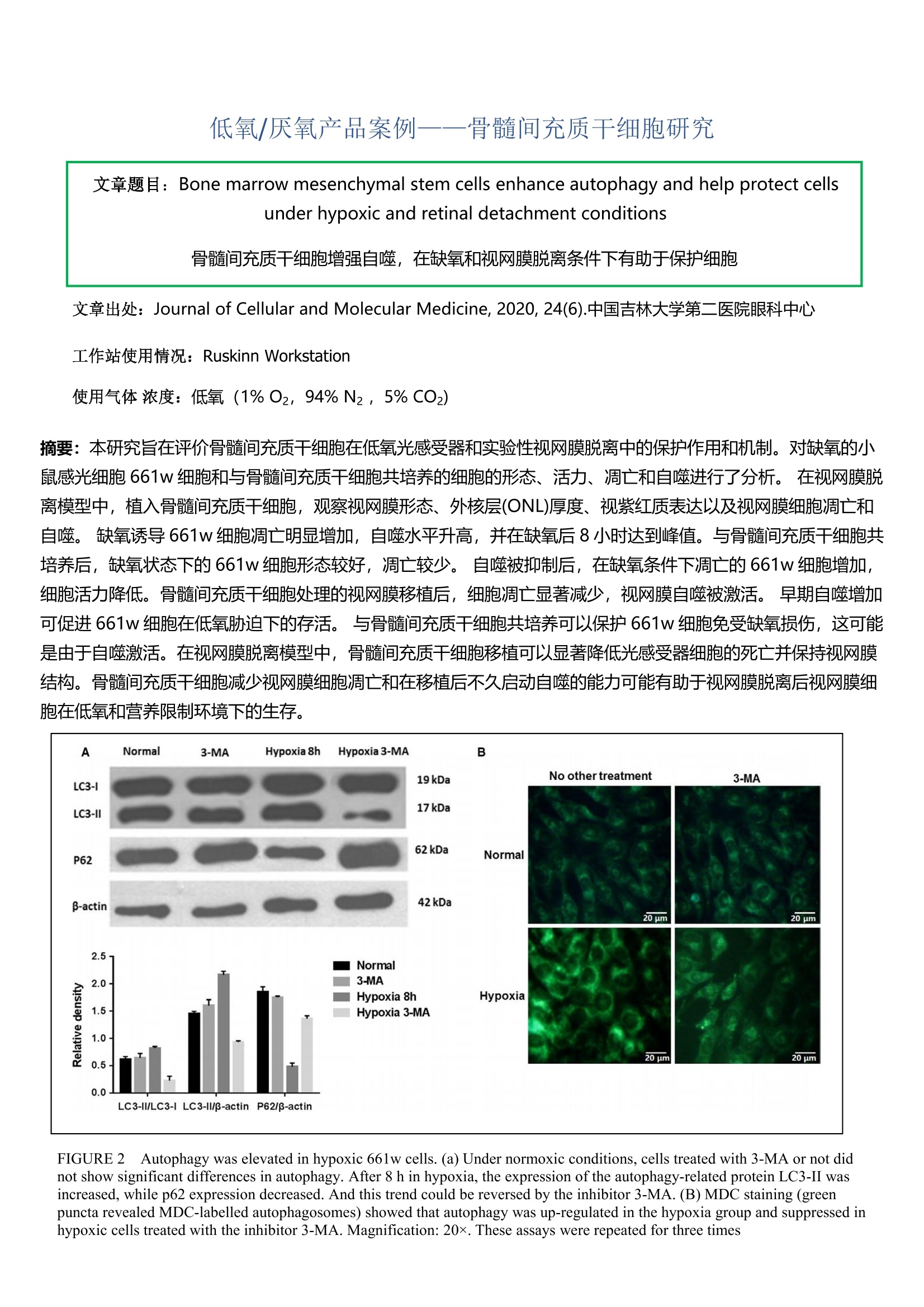
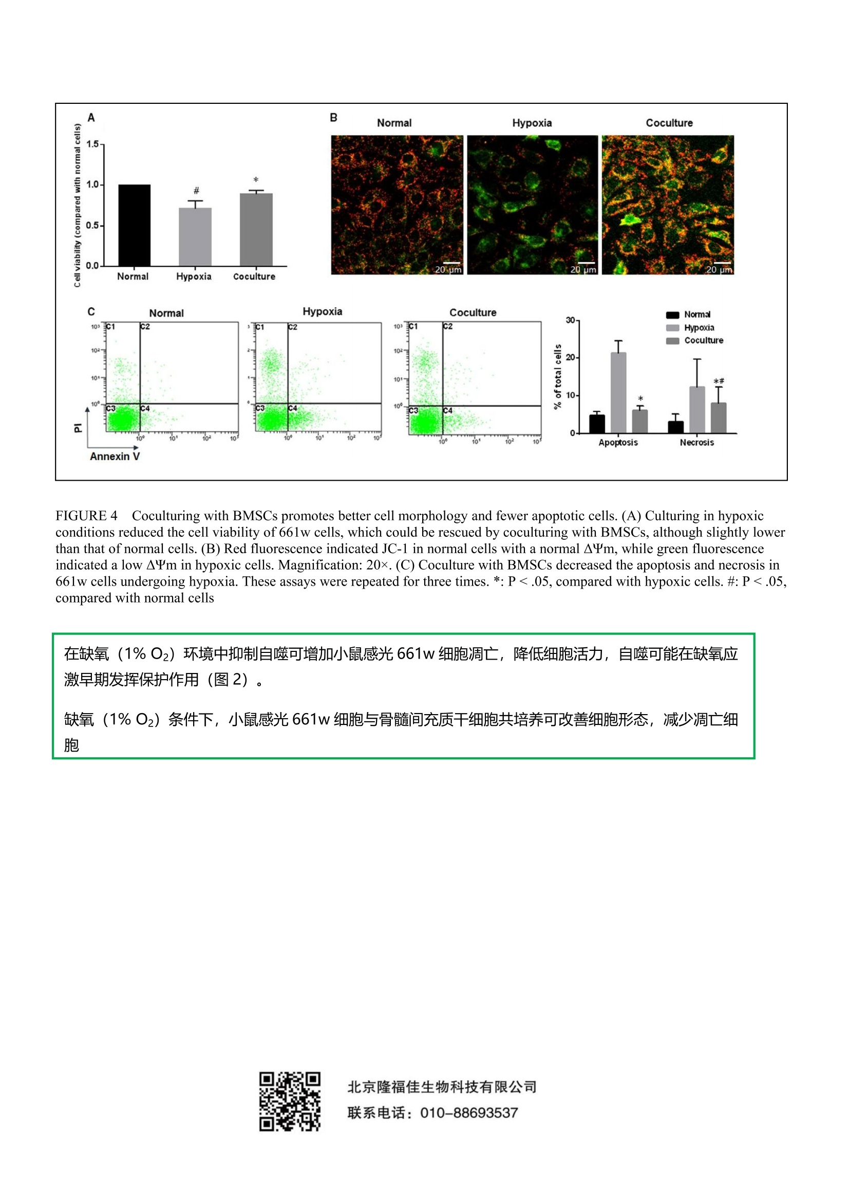
低氧/厌氧产品案例――骨髓间充质干细胞研究 文章题目: Bone marrow mesenchymal stem cells enhance autophagy and help protect cellsunder hypoxic and retinal detachment conditions 骨髓间充质干细胞增强自噬,在缺氧和视网膜脱离条件下有助于保护细胞 文章出处: Journal of Cellular and Molecular Medicine, 2020, 24(6).中国吉林大学第二医院眼科中心 工作站使用情况:Ruskinn Workstation 使用气体浓度:低氧(1%02, 94%N2, 5% CO2) 摘要:本研究旨在评价骨髓间充质干细胞在低氧光感受器和实验性视网膜脱离中的保护作用和机制。对缺氧的小鼠感光细胞 661w 细胞和与骨髓间充质干细胞共培养的细胞的形态、活力、凋亡和自噬进行了分析。在视网膜脱离模型中,植入骨髓间充质干细胞,观察视网膜形态、外核层(ONL)厚度、视紫红质表达以及视网膜细胞凋亡和自噬。缺氧诱导661w细胞凋亡明显增加,自噬水平升高,并在缺氧后8小时达到峰值。与骨髓间充质干细胞共培养后,缺氧状态下的661w细胞形态较好,凋亡较少。自噬被抑制后,在缺氧条件下凋亡的661w细胞增加,细胞活力降低。骨髓间充质干细胞处理的视网膜移植后,细胞凋亡显著减少,视网膜自噬被激活。早期自噬增加可促进661w细胞在低氧胁迫下的存活。与骨髓间充质干细胞共培养可以保护661w细胞免受缺氧损伤,这可能是由于自噬激活。在视网膜脱离模型中,骨髓间充质干细胞移植可以显著降低光感受器细胞的死亡并保持视网膜结构。骨髓间充质干细胞减少视网膜细胞凋亡和在移植后不久启动自噬的能力可能有助于视网膜脱离后视网膜细胞在低氧和营养限制环境下的生存。 FIGURE22AAutophagy was elevated in hypoxic 661w cells.(a) Under normoxic conditions, cells treated with 3-MA or not didnot show significant differences in autophagy. After 8 h in hypoxia, the expression of the autophagy-related protein LC3-II wasincreased, while p62 expression decreased. And this trend could be reversed by the inhibitor 3-MA. (B) MDC staining (greenpuncta revealed MDC-labelled autophagosomes) showed that autophagy was up-regulated in the hypoxia group and suppressed inhypoxic cells treated with the inhibitor 3-MA. Magnification: 20x. These assays were repeated for three times FIGURE 4 Coculturing with BMSCs promotes better cell morphology and fewer apoptotic cells. (A) Culturing in hypoxicconditions reduced the cell viability of 661w cells, which could be rescued by coculturing with BMSCs, although slightly lowerthan that of normal cells. (B) Red fluorescence indicated JC-1 in normal cells with a normal AYm, while green fluorescenceindicated a low AYm in hypoxic cells. Magnification: 20x. (C) Coculture with BMSCs decreased the apoptosis and necrosis in661w cells undergoing hypoxia. These assays were repeated for three times.*:P<.05, compared with hypoxic cells. #: P <.05,compared with normal cells 在缺氧(1%02)环境中抑制自噬可增加小鼠感光661w细胞凋亡,降低细胞活力,自噬可能在缺氧应激早期发挥保护作用(图2)。 缺氧(1%02)条件下,小鼠感光661w 细胞与骨髓间充质干细胞共培养可改善细胞形态,减少凋亡细胞 北京隆福佳生物科技有限公司 联系电话:010-88693537 文章题目:Bone marrow mesenchymal stem cells enhance autophagy and help protect cells under hypoxic and retinal detachment conditions骨髓间充质干细胞增强自噬,在缺氧和视网膜脱离条件下有助于保护细胞文章出处:Journal of Cellular and Molecular Medicine, 2020, 24(6).中国吉林大学第二医院眼科中心工作站使用情况:Ruskinn Workstation使用气体浓度:低氧(1% O2,94% N2 ,5% CO2)摘要:本研究旨在评价骨髓间充质干细胞在低氧光感受器和实验性视网膜脱离中的保护作用和机制。对缺氧的小鼠感光细胞661w 细胞和与骨髓间充质干细胞共培养的细胞的形态、活力、凋亡和自噬进行了分析。在视网膜脱离模型中,植入骨髓间充质干细胞,观察视网膜形态、外核层(ONL)厚度、视紫红质表达以及视网膜细胞凋亡和自噬。缺氧诱导661w 细胞凋亡明显增加,自噬水平升高,并在缺氧后8 小时达到峰值。与骨髓间充质干细胞共培养后,缺氧状态下的661w 细胞形态较好,凋亡较少。自噬被抑制后,在缺氧条件下凋亡的661w 细胞增加,细胞活力降低。骨髓间充质干细胞处理的视网膜移植后,细胞凋亡显著减少,视网膜自噬被激活。早期自噬增加可促进661w 细胞在低氧胁迫下的存活。与骨髓间充质干细胞共培养可以保护661w 细胞免受缺氧损伤,这可能是由于自噬激活。在视网膜脱离模型中,骨髓间充质干细胞移植可以显著降低光感受器细胞的死亡并保持视网膜结构。骨髓间充质干细胞减少视网膜细胞凋亡和在移植后不久启动自噬的能力可能有助于视网膜脱离后视网膜细胞在低氧和营养限制环境下的生存。FIGURE 2 Autophagy was elevated in hypoxic 661w cells. (a) Under normoxic conditions, cells treated with 3-MA or not did not show significant differences in autophagy. After 8 h in hypoxia, the expression of the autophagy-related protein LC3-II was increased, while p62 expression decreased. And this trend could be reversed by the inhibitor 3-MA. (B) MDC staining (green puncta revealed MDC-labelled autophagosomes) showed that autophagy was up-regulated in the hypoxia group and suppressed in hypoxic cells treated with the inhibitor 3-MA. Magnification: 20×. These assays were repeated for three times.FIGURE 4 Coculturing with BMSCs promotes better cell morphology and fewer apoptotic cells. (A) Culturing in hypoxic conditions reduced the cell viability of 661w cells, which could be rescued by coculturing with BMSCs, although slightly lower than that of normal cells. (B) Red fluorescence indicated JC-1 in normal cells with a normal ΔΨm, while green fluorescence indicated a low ΔΨm in hypoxic cells. Magnification: 20×. (C) Coculture with BMSCs decreased the apoptosis and necrosis in 661w cells undergoing hypoxia. These assays were repeated for three times. *: P < .05, compared with hypoxic cells. #: P < .05, compared with normal cells.在缺氧(1% O2)环境中抑制自噬可增加小鼠感光661w 细胞凋亡,降低细胞活力,自噬可能在缺氧应激早期发挥保护作用(图2)。缺氧(1% O2)条件下,小鼠感光661w 细胞与骨髓间充质干细胞共培养可改善细胞形态,减少凋亡细胞。
确定
还剩1页未读,是否继续阅读?

产品配置单
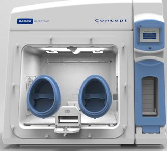
北京隆福佳生物科技有限公司为您提供《骨髓间充质干细胞中增强自噬检测方案(厌氧培养箱)》,该方案主要用于其他中其他检测,参考标准--,《骨髓间充质干细胞中增强自噬检测方案(厌氧培养箱)》用到的仪器有Concept 400M低氧/厌氧工作站(低氧/厌氧培养箱)
相关方案
更多
该厂商其他方案
更多