


方案详情
文
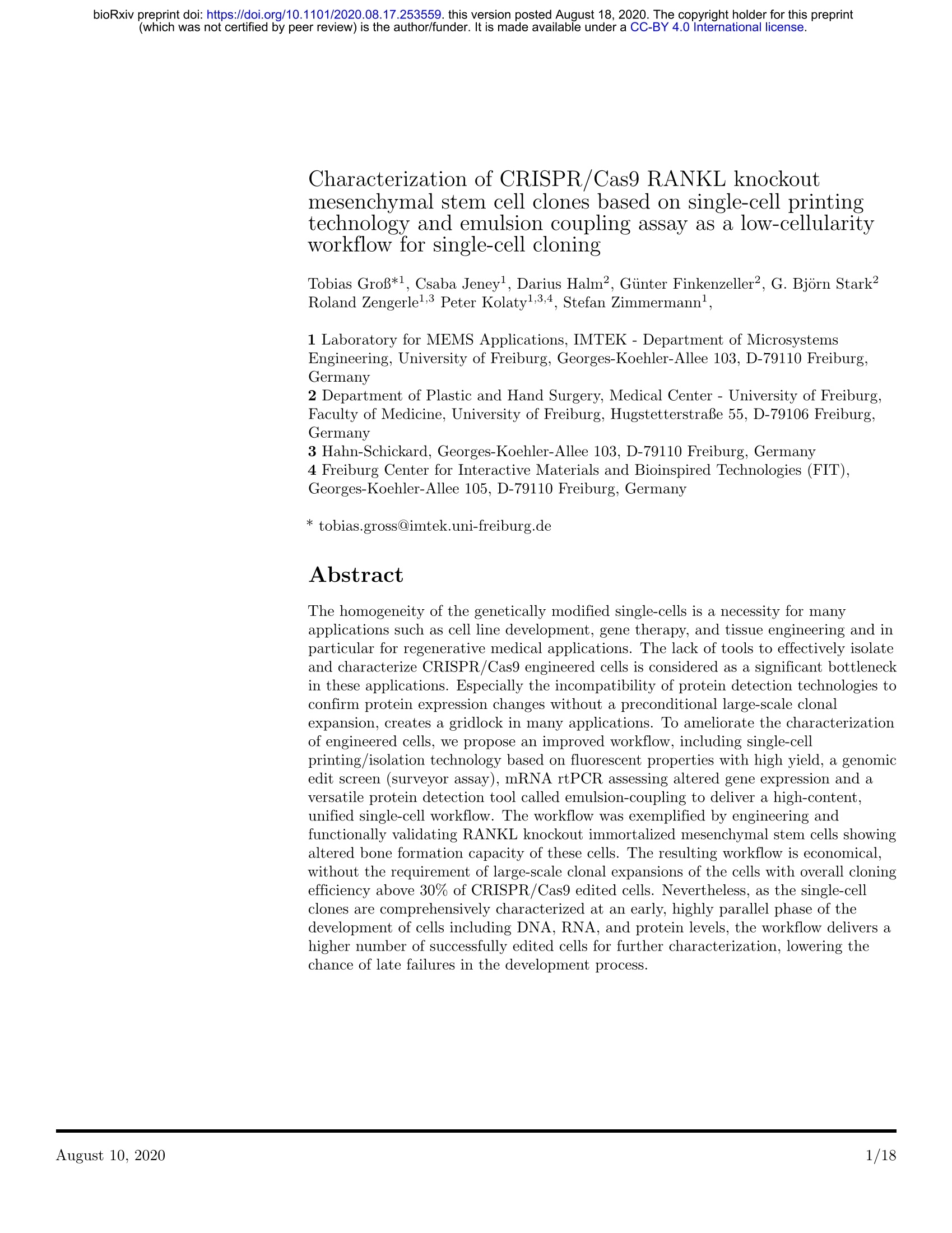
CloneSelect单细胞分离系统(CloneSelect Single Cell Printer)采用类似喷墨打印的技术,柔和地产生包裹细胞的液滴无接触地直接分配到微孔板中(图1)。同时,该系统借助智能图像分析,确认细胞数目,分析细胞的形态(大小和圆度)及荧光强度,联动的真空装置将不符合要求的液滴(如空液滴或者含多个细胞的液滴)直接吸走,而符合要求的细胞液滴则分配至微孔板,从而实现对单个细胞的分选和接种。单细胞分离系统是一种可比移液器操作的柔和的单细胞分离技术,而流式分选由于高的液体剪切力和电压的影响,会降低敏感细胞和部分受损细胞(如电转后的细胞)的成克隆率,因此单细胞分离系统更适用于基因工程细胞的克隆,维持细胞活力,并提供直接的单克隆性图像证据。
方案详情

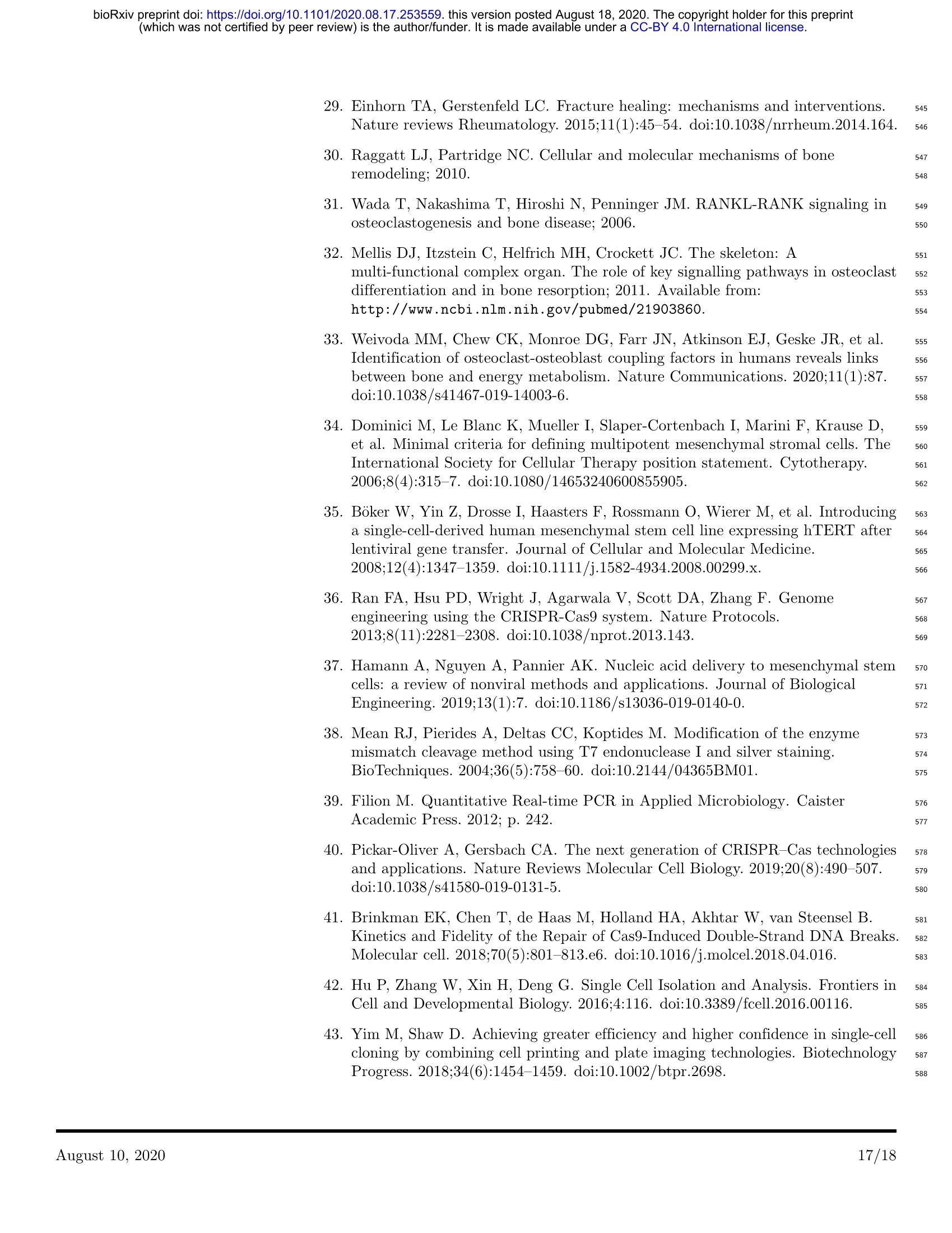
bioRxiv preprint doi:https://doi.org/10.1101/2020.08.17.253559. this version posted August 18, 2020. The copyright holder for this preprint(which was not certified by peer review) is the author/funder. It is made available under a CC-BY 4.0 International license. bioRxiv preprint doi:https://doi.org/10.1101/2020.08.17.253559. this version posted August 18, 2020. The copyright holder for this preprint Characterization of CRISPR/Cas9 RANKL knockoutmesenchymal stem cell clones based on single-cell printingtechnology and emulsion coupling assay as a low-cellularityworkflow for single-cell cloning Tobias GroB*1, Csaba Jeney, Darius Halm2, Gunter Finkenzeller2, G. Bjorn Stark2Roland Zengerlel,3Peter Kolaty1,3,4, Stefan Zimmermannl, 1 Laboratory for MEMS Applications, IMTEK - Department of MicrosystemsEngineering,University of Freiburg, Georges-Koehler-Allee 103, D-79110 Freiburg,Germany 2 Department of Plastic and Hand Surgery, Medical Center -University of Freiburg,Faculty of Medicine, University of Freiburg, HugstetterstraBe 55, D-79106 Freiburg,Germany 3 Hahn-Schickard, Georges-Koehler-Allee 103, D-79110 Freiburg, Germany4 Freiburg Center for Interactive Materials and Bioinspired Technologies (FIT),Georges-Koehler-Allee 105, D-79110 Freiburg, Germany * tobias.gross@imtek.uni-freiburg.de Abstract The homogeneity of the genetically modified single-cells is a necessity for manyapplications such as cell line development, gene therapy, and tissue engineering and inparticular for regenerative medical applications. The lack of tools to effectively isolateand characterize CRISPR/Cas9 engineered cells is considered as a significant bottleneckin these applications. Especially the incompatibility of protein detection technologies toconfirm protein expression changes without a preconditional large-scale clonalexpansion, creates a gridlock in many applications. To ameliorate the characterizationof engineered cells, we propose an improved workflow, including single-cellprinting/isolation technology based on fluorescent properties with high yield, a genomicedit screen (surveyor assay), mRNA rtPCR assessing altered gene expression and aversatile protein detection tool called emulsion-coupling to deliver a high-content,unified single-cell workflow. The workflow was exemplified by engineering andfunctionally validating RANKL knockout immortalized mesenchymal stem cells showingaltered bone formation capacity of these cells. The resulting workflow is economical,without the requirement of large-scale clonal expansions of the cells with overall cloningefficiency above 30% of CRISPR/Cas9 edited cells. Nevertheless, as the single-cellclones are comprehensively characterized at an early, highly parallel phase of thedevelopment of cells including DNA, RNA, and protein levels, the workflow delivers ahigher number of successfully edited cells for further characterization, lowering thechance of late failures in the development process. bioRxiv preprint doi:https://doi.org/10.1101/2020.08.17.253559. this version posted August 18, 2020. The copyright holder for this preprint (which was not certified by peer review) is the author/funder. It is made available under a CC-BY 4.0 International license. Author summary I completed my undergraduate degree in biochemistry at the University of Ulm andfinished my master’s degree in pharmaceutical biotechnology at the University of Ulmand University of applied science of Biberach with a focus on biotechnology, toxicologyand molecular biology. For my master thesis, I went to the University of Freiburg to thedepartment of microsystems engineering, where I developed a novel workflow for cellline development. I stayed at the institute for my doctorate, but changed my scientificfocus to the development of the emulsion coupling technology, which is a powerful toolfor the quantitative and highly parallel measurement of protein and protein interactions.I am generally interested in being involved in the development of innovative molecularbiological methods that can be used to gain new insights about biological issues. I amparticularly curious to unravel the complex and often poorly understood proteininteraction pathways that are the cornerstone of understanding cellular functionalityand are a fundamental necessity to describe life mechanistically. (which was not certified by peer review) is the author/funder. It is made available under a CC-BY 4.0 International license. Introduction There is a high demand for well-characterized genetically engineered single-cellclones [1]. The assurance of their clonality and lineage traceability are important notonly for pharmaceutical but also for cell therapeutic applications particular forregenerative medical uses [2], which is also enforced by the regulatory requirements ofthe European Medicine Agency (EMA) and the Food and Drug Administration(FDA) [3,4]. This work aims to provide an improved, more parallel workflow, withouttime-consuming clonal expansion, to generate deeply characterized single-cell clones thatcan meet these quality requirements. 2 5 6 7 8 Most genetic engineering methods such as CRISPR/Cas9 are error-prone, generating 10a non-homogeneous population of cells by failing to introduce the engineered changes 11correctly, having off-targets, monoallelic modifications, and many non-edited cells [5], 12rendering the clonal isolation of the cells and the characterization of the clones 13mandatory before their use. Nevertheless, as the genomically correct modification does 14not ensure the intended gene expression changes, the validation of the requirements 15necessitates a high-content characterization at genomic,mRNA expression, and also 16protein level. To fulfill these analytical needs a battery of technologies is applied, which 17introduce their own, in many cases, disparate requirements. As a consequence, the 18current approaches frequently include the costly, failure-prone, and time-consuming 19expansion of the cells solely to provide material for the subsequent analytical methods. 20This is especially true for protein analytic technologies, as they are in regard to most 21frequently used methods, and in contrast to genomic and RNA expression technologies, 22not molecularly sensitive. While genomic and RNA expression detection technologies 23can use even a single cell of sample for their analysis [6], protein analytics need several 24magnitude larger sample amounts. The available classic methods such as mass flow 25cytometry, mass spectrometry, ELISA, or Western Blot often require a large number of 26cell material in order to detect the targeted protein [7-10]. However, new protein 27analytical technologies are emerging such as proximity ligation assay [11], proximity 28extension assay [12], or single-cell mass spectrometry [13]. 29 Recently, single-cell printing technology (SCP) is emerging from others [14, 15] as agentle, low-cost and highly controlled technology, applicable for a wide array of specificcell-cloning applications ranged from the 0.8 um prokaryotes to 100 um plantcells [16,17]. It is based on inject-jet-like technologies generating on-demand free-flyingmicrodroplets encapsulating cells to deposit them on variable substrates using anon-contact dispensing process. The technology is described in more detail by Gross etal. 2013. 30 31 32 33 34 35 36 SCP can be considered as a very gentle single-cell isolation technology that is 37comparable to manual pipetting in terms of the applied shear stress [18]. SCP is also 38superior to fluorescence-associated cell sorting (FACS), which is also frequently used for 39single-cell isolation, as FACS has a negative effect on cloning efficiency of sensitive or 40partly-damaged cells (e.g. by transfection) by applying high shear forces and 41electrostatic charging of cells [15]. SCP is especially well-suited for cloning of engineered 42cells, besides preserving their viability, it provides direct proof of single-cell clonality 43(e.g. delivers trustable records of each printed cell), has both native and fluorescent 44sorting capabilities, and can be coupled to single-cell analytical pipelines. Recently, SCP 45has become a frequent choice for single-cell isolation purposes in different workflows 46including subsequent analytics [19]. 47 As discussed above, genomic and transcriptomic assays usually do not posesignificant issues but the mandatory protein analytics are a central challenge in cloningworkflows. Albeit the recent developments of sensitive protein assays and their widevariety of applications further improvement is still necessary. An ideal assay has ahomogenous measurement principle (e.g. no washing steps),can be used for sample 48 49 50 51 52 bioRxiv preprint doi:https://doi.org/10.1101/2020.08.17.253559. this version posted August 18, 2020. The copyright holder for this preprint sizes with very low cell numbers down to a few dozen cells, and yet provides highlysensitive and quantitative detection of many proteins parallel, including proteinpost-translational modifications, and protein-protein interactions. Recently, emulsioncoupling [20] promises to fulfill these requirements. 53 Emulsion coupling is based on a digital method that is similar to the droplet digitalPCR (ddPCR) [21,22] and detects the partitioning differences of two DNAoligonucleotide labeled antibodies per target in the presence of target protein comparedto their free, unbound distribution in ddPCR emulsion. This assay is homogenous,molecularly sensitive, quantitative, and due to its two antibody-principle, it is highlyspecific and can be read by sequencing (e.g. by next-generation sequencing) in a highlyparallel way. Its low sample requirement and parallelity make it an ideal candidate toestablish a cell cloning workflow with minimum clonal expansion. 5455565758596061626364To provide an experimental proof of concept, immortalized mesenchymal stem cells 65have been genetically engineered to improve their bone-forming capacity for potential 66regenerative medical applications. For this, Tumor Necrosis Factor Superfamily Member 6711 (TNFSF11) was knocked out as suggested among others by Walsh and Choi 682014 [23-25], which encodes the receptor activator for nuclear factor kappa B ligand 69(RANKL) [26]. RANKL has been shown to play a crucial role in bone homeostasis by 70orchestrating the balance between bone-generating osteoblasts and bone-degrading 71osteoclasts [27-29]via the so-called OPG/RANKL/RANK pathway [30]. Briefly, 72RANKL is expressed in bone tissue by mesenchymal stem cells (MSCs), osteoblasts, and 73T-cells, among others [31]. In the presence of RANKL, the receptor activator for nuclear 74factor kappa B (RANK) is activated which stimulates pre-osteoclasts to differentiate 75into osteoclasts which in turn degrade bone 32,33]. For bone formation, MSCs 76differentiate into osteoblasts and deposit calcified structures 34]. By TNFSF11 77knockout, the genetically modified MSCs and their progenitors can no longer recruit 78osteoclasts and as a consequence, the bone formation at the side of their implantation in 79a regenerative medical application could be potentially improved. 8081828384 An improved workflow was provided including SCP technology with fluorescentsorting, and limited clone expansion to enable genomic edit screen (surveyor assay),mRNA RT-PCR assessing altered gene expression, and emulsion coupling to deliver ahigh-content,unified single-cell cloning workflow. Materials and methods 85 Cell culture 86 Immortalized MSCs were kindly provided by Prof. Dr. Matthias Schieker from the 87Laboratory of Experimental Surgery and Regenerative Medicine of the 88 Ludwig-Maximilians-University of Munich. They were immortalized by retroviral 89transduction of human telomerase reverse transcriptase (hTERT MSC) [35]. The cells 90were cultivated in NuncEasYFlaskNunclonDelta Surface (Thermo Scientific) 91using the cell culture medium MEM alpha Medium (1x) + GlutaMax"-I (Gibco), 9210%FBS (Gibco) and 1% Pen-Strep (Gibco) at 37°℃/5% CO2. 93 Determination of the cumulative population doublings 94 The population doubling (PD) was determined by tracking the cell number of the 95initially seeded cells (y) and the cell yield during cell passaging(x). The PD was 96calculated by the following formula: PD=(ln(x)-ln(y))/ln(2). The cumulative 97population doubling (CPD) is the total number of times the cells in a given population 98have doubled in culture, which was plotted against the days after single-cell isolation. 99 Plasmids 100 The RANKL specific gRNA construct (pNV-RANKL/KO) based on the parenteral 101 gRNA-Cas9-2A-GFP vector, which is transiently expressing gRNA and Cas9 and 102 enables GFP selection of transfected MSCs, was purchased from abm (Richmond, 103Canada). Three vectors were purchased with the following gRNA sequences: 104pNV-RANKL1/KO-5'-CAGGAATTACAACATATCGT-3’(472221110290), 105pNV-RANKL2/KO-5'-CAGCGATGGTGGATGGCTCA-3'(472221110390), 106pNV-RANKL3/KO-5'-TTAATAGTGAGATGAGCAAA-3'(472221110490). pmaxGFP 107(purchased from Lonza, Human MSC NucleofectorR Kit) was used to control 108transfection efficiency. Plasmids were propagated by transforming Escherichia Coli 109(Mix&Go! Competent Cells - DH5 Alpha, Zymo Research) using standard procedures. 110 The plasmids were purified using the ZymoPURE II Plasmid Midiprep Kit (Zymo 111 Research) according to the manufacturer’s instructions. The plasmid concentration was 112measured with the NanoDropT One. The plasmids were stored at - 20°C. 113 Transfection 114 Sub- confluent hTERT MSCs were trypsinized,harvested, and washed by PBS using 115standard procedures. For nucleofection of hTERT MSC cells, cells were resuspended in 116100 ul Human MSC Nucleofector Solution (Human MSC NucleofectorR Kit-Lonza) 117and the transfection was carried out according to the manufacturer’s instructions using 118different amount of gRNA plasmids or pmaxGFP plasmid. The transfected cells were 119incubated at 37°C/5% CO2 for 24 h before further steps. Transfection efficiencies were 120determined using the data obtained during single-cell printing. GFP expressing cells 121were set relative to the total count of detected cells. 122 Single-cell printing and single-cell cloning 123 printed into wells of a 96 well-plate (flat bottom clear polystyrene, Greiner) containing 128200 ul cell culture medium (see cell culture). After printing, the printed cells were 129detected with the high-resolution imager NyOne (SynenTec) either in single-cell colony 130(SCC) or in fluorescence-activated SCC (FASCC) mode. The single-cell printing 131efficiency was determined based on Gross et al. 2013. Briefly, images that were taken of 132the printed cells during the printing process were manually analyzed for single, multiple, 133void, or uncertain printing events. The percentage of each event was calculated relative 134to the total printing events. The single-cells cloning efficiency was accessed by the 135high-resolution imager NyOne (SynenTec) using its confluence mode. Cloning 136efficiency was determined by calculating the percentage of the wells with cell colonies 137relative to the total count of printed wells with single-cells. 138 Genome editing detection 139 Genomic DNA was extracted from 8.0x10 hTERT MSCs with the Quick-DNA 140Microprep Plus Kit (Zymo Research) and then analyzed for indel mutations by the 141surveyor assay using the Alt-RGenome Editing Detection kit (IDT) according to the 142manufacturer’s instructions. Briefly, the genomic DNA was extracted from the samples 143and the loci of the edited sites were amplified by PCR of both. Amplifications were 144carried out using the HotStarTaq Plus Master Mix Kit (Qiagen) according to the 145manufacturer's instructions. Unmodified hTERT MSC reference with CRISPR edited 146DNA was combined to form heteroduplexes in a 1:1 ratio, 5 ul unmodified hTERT MSC 147reference with 5 ul CRISPR edited DNA. The heteroduplexes were digested according 148to the Alt-R Genome Editing Detection Kit. Controls A and B from the Alt-R 149Genome Editing Detection Kit were used and treated in the same way. 4 ul of the 150digested DNA was mixed with 2 pl1:100 in DMSO diluted GelRedM (Biotium) dye and 151loaded onto a 1% agarose gel and as a ladder the 100 bp plus DNA ladder from VWR 152was used. Gel images were taken by the camera system INGenius (Syngene). The 153forward primer 5'-AAGTTCTGCGGCCCAGTTTA-3'and reverse primer 1545'-AGGGAGAGAAAGGAACCTCTG-3'were used. PCR conditions having a hot-start 155at 95℃ for 5 min, with cycling conditions at 95°℃ for 30 sec, 30 sec annealing 156temperature at 64°C, and with an extension time of 1.5 min at 72℃ and 40 cycle 157rounds with a final extension time at 72℃ for 10 min. Control A/B shows three bands 158at 690 bp, 463 bp, and 256 bp for digested heteroduplexes, control A for a homoduplex 159at 690 bp, genomic DNA from unmodified hTERT MSCs at 1850 bp and the negative 160 control (-) no signal. 161 Real-time quantitative PCR 162 The gene expression level of TNFSF11 in hTERT MSCs and the RANKL-KO hTERT 163 MSCs were determined using qPCR. Briefly, total RNA was isolated from 5×10cells 164with the AllPrep@ DNA/RNA/Protein Mini Kit (Qiagen) according to the 165manufacturer’s instructions. Two assays were used simultaneously with a VIC-probe 169located at the 5'end of the TNFSF11 transcript (Hs00243519_m1 TaqMan assay) and a 167FAM-probe located at the 3’end of the TNFSF11 transcript (Hs00243522_m1 TaqMan 168assay). The qPCR was performed using the TaqManRNA-to-CT 1-Step kit 169(Thermo Fischer Scientific) and was run in a RotorGene-6000-2-plex (Qiagen). PCR 170conditions having reverse transcription at 48°℃ for 15 min, hot-start at 95℃ for 10 min, 171with cycling conditions starting with a denaturation step at 95C for 15 sec, 172 anneal/extend step at 60℃ for 1 min. The results were analyzed with the Rotor-GeneQ Series Software from Qiagen. 173 174 Emulsion Coupling Assay 10 hTERT MSCs, and different RANKL-KO (see section Plasmids) hTERT MSCs 176 were harvested and lysed and processed according to the emulsion coupling protocol 177described by Karakus et al. 2019 and is briefly described below. Two anti-RANKL 178antibodies, RANKL-G1 (Santa Cruz Biotechnology) FAM signal, anti-CD254 179(Biolengend) VIC signal, were used. The oligonucleotide labeling of the antibodies was 180carried out according to Karakus et al. 2019. Lysed cells were incubated with the 181antibodies overnight and diluted 100,000-fold. Emulsification, ddPCR, and readout 182were performed using the QX200Droplet DiDgi1talPCR System (Bio-Rad) following 183standard ddPCR protocol. As a negative control, the antibodies were mixed without 184antigen and processed in the same way as the samples (antibody-binding-control, ABC). 185The amount of protein RANKL was measured by detecting the partitioning differences 186induced by concurrent binding of the antibodies to RANKL. The measurements were 187normalized against the ABC control, which defines the zero level detection by the ABC 188 signal. 189 Osteogenic differentiation 190 104 cells of the CRISPR-edited RANKL knockout cell clones and unmodified hTERT 191MSCs were seeded in 6-well plates. They were incubated in 2 ml of osteogenic 192differentiation medium (DMEM low glucose (Gibco), 10%FBS, 1%PenStrep, 100 nM 193dexamethasone, 50 ug/ml ascorbate, 10 mM B-glycerophosphate) for 7, 14 and 21 days. 194In order to stain calcified deposits, 2 gof Alizarin Red (Sigma-Aldrich) was dissolved in 195100 ml distilled water and the pH was adjusted to 4.5 -5 by NaOH and the solution 196was filtered. Before staining, the culture medium was removed from the cells and they 197were washed 2 times with PBS. The cells were then fixed with 4% formalin in PBS for 19810 minutes and washed 2 times with PBS. For staining, 2 ml Alizarin Red staining 199solution was added to the wells, and the cells were stained in the dark for 45 minutes. 200The staining solution was removed and the cells were carefully washed 5 times with 201PBS. Images of the stained cells were captured with the microscope DMil (Leica). 202 Results and Discussion Experimental design 204 Working on the generation of genetically engineered cells raises a number of problems, 205among others the generation of well-characterized clones and their assurance and 206documentation of clonality. We propose a practicable workflow for single-cell cloning 207addressing these problems which additionally provides low-cellularity analytics. The 208individual steps of the improved workflow are displayed in Fig1. To characterize a large 209number of clones in economical timescales without excessive clonal expansion, PCR 210based technologies are available. However, such technologies are highly limited in 211protein analytics. We devised a technological solution to overcome this limitation: (i) 212modifications at the genomic level were detected using a surveyor assay, (ii) 213transcription levels of target genes were checked by RT-PCR and (iii) target protein 214expression by the clones was measured by emulsion coupling. All these assays enable 215the characterization of the clones from a few hundred to thousand cells as sample 216amount (low-demand assay -LDA), fostering low clonal expansion and high throughput 217characterization, but still providing high-content information about the clones, 218facilitating successful functional assays of the cherry-picked clones. This workflow is 219compatible with industrial requirements, highly automatable, and can easily be 220transferred to other cell types and genetic targets. For a proof of concept, the workflow 221was applied on hTERT MSCs which were TNFSF11 edited using the CRISPR/Cas9 222system [36], to demonstrate the efficient single-cell handling and sorting of genetically 223modified cells using SCP as well as accessing the performance of the LDAs in this 224environment. 225 Fig 1. Improved workflow to establish and characterize CRISPR/Cas9edited cells with minimal clonal expansion. gRNAs transfected cells wereisolated and sorted by SCP detecting GFP signal. To characterize the introducedgenetic changes, RNA, and protein expressions, a set of assays requiring a low numberof cells was proposed: (i) surveyor assay (DNA edits), (ii) RT-PCR(RNA expression)and (iii) emulsion coupling (protein expression). The selected clones are ready forfurther confirmatory assays (iv). Arrows indicate gRNAs, red crosses symbolizeintroduced genetic changes. CRISPR/Cas9 gene editing and single-cell cloning hTERT MSCs were transfected with the pNV-RANKL/KO plasmids, CRISPR/Cas9 227knockout vectors, with a GFP marker gene containing different gRNAs directed against 228TNFSF11 (see Material and Methods). In order to sort transfected single-cells from the 229untransfected ones, the SCP technology with fluorescent sorting (cytena, f.sightM) was 230applied by detecting and sorting cells by the GFP signal of the transfected constructs. 231In Fig 2A, exemplary five images of the isolation process of a transfected and GFP 232expressing hTERT MSC are shown. As a control, hTERT MSCs were transfected with 233pmaxGFP to evaluate the efficiency of printing, sorting, and cloning and to optimize 234the process. hTERT MSCs were transfected with pNV-RANKL/KO gRNA variants and 235printed and sorted according to the determined parameters with regard to size, 236roundness and laser intensity, exposure time, and fluorescent threshold. Although the 237hTERT MSCs were transfected with different transfection efficiencies, pmaxGFP had 238an efficiency of 61% and pNV-RANK/KO only 6%, and additionally, pNV-RANK/KO 239transfection induced significantly more dead cells, at the same time a cloning efficiency 240of 30% was achieved with the transfected cells. The differences in transfection efficiency 241 can be explained by the smaller size of the pmaxGFP control vector, which is 242approximately 3.5 kb compared to 12 kb of the pNV-RANKL/KO plasmid [37]. The 243pmaxGFP plasmid was designed to have high-intensity GFP expression while the SFFV 244promoter in the pNV-RANKL/KO vector transcribed a Cas9-GFP multicistronic 245transcription unit, resulting in a weaker signal.We also noted a minimal difference in 246the cloning efficiency of transfected hTERT MSCs (31.3 ±8.0%) and non-transfected 247hTERT MSCs (39.6 士15.6%) indicating the efficacy of the sorting/printing process is 248gentle and has a minimum capacity to introduce systematic biases(see Fig 2G). 249 Fig 2. Comparison of the single-cell printing and cloning efficiency ofpmaxGFP and pNV-RANKL/KO transfected hTERT MSCs. hTERT MSCswere transfected with either pmaxGFP (transfection efficiency 61%) orpNV-RANKL/KO (transfection efficiency 6%). Two days post-transfection, GFPexpressing single-cells were isolated by SCP (see details below). In (A) the SCP(f.sight ) cartridge ejection area is recorded during the printing process, showing afluorescent single-cell before (01-03), at (04), and after (05) ejection. The SCPalgorithm intercepts and deflects cells that are not qualifying the preset parameters(size threshold was set from 15-30 um, roundness from 0.6-1, where 1 reflects theperfectly circular object). In (B) and (D) the roundness is plotted against cell diameterand in (C) and (E) the relative fluorescence intensity unit (RFU) is plotted against celldiameter - all detected events are in gray. The red dots represent printed single-cellsqualified to the preset parameters (see below). The dotted orange line represents thethresholds of parameters for selecting single cells. (F) The printed single-cells wereevaluated for clonality (n= 451 transfected printed cells). The recorded image series ofeach qualified printing process was evaluated manually, categorizing single, multiple-cell,uncertain, or void printing events. (G) After 2 weeks colonies obtained from single cellswere counted and thereby the cloning efficiency determined (n=227 transfected printedcells, n=650 non-transfected printed cells). The single-cell printing parameters appliedfor pmaxGFP transfected hTERT MSCs:1laser intensity of 20 - 40%, an exposure timeof 15 -30 ms and a fluorescence threshold of 30 -250 RFU while for pNV-RANKL/KOtransfected hTERT MSCs laser intensity of 90-95%, exposure time of 80 - 120 ms and afluorescence threshold of 50- 250 RFU. In Fig 2B to E), all recorded events (e.g. debris or living and dead cells) during SCP 250were plotted against their parameters diameter, roundness, or RFU from one consecutive 251experiment of either pmaxGFP or pNV-RANKL/KO transfected hTERT MSCs. The 252cells were sorted in a size range of 15 to 35 um and roundness of 0.6 to 1, where 1 253reflects a perfectly circular object. In Fig 2B and C, the diameter against roundness is 254plotted while in Fig 2C and D the RFU is plotted against the diameter. The majority of 255the recorded events show RFU values below 30 but strong population showed an RFU 256above 30, representing the GFP expressing MSCs with a potential RANKL knockout. 257The dots marked in red indicate sorted events. The printed cells were selected from an 258RFU above 30 for pmaxGFP transfected hTERT-MSCs while pNV-RANKL/KO cells 259were selected from an RFU above 50. In addition, the series of images recorded during 260the printing process suggest that only cells have been sorted (see exemplarily Fig. 2 A). 261The selection threshold values are indicated in the graphs by orange dashed lines. 262 The single-cells were individually sorted and printed in 96-well plates, filled with 263pre-warmed cell culture medium, and evaluated manually. The first line of analysis is 264based on the series of images that were taken during the printing process and analyzed 265manually, whether fluorescent single-cells were successfully sorted and printed or 266whether multiple, an uncertain amount, no cells or non-fluorescent cells were delivered 267into the wells. This analysis gives the fluorescent single-cell printing efficiency which is 268shown in Fig 2F. The combined fluorescent single-cell printing efficiency of all 269 bioRxiv preprint doi:https://doi.org/10.1101/2020.08.17.253559. this version posted August 18, 2020. The copyright holder for this preprint experiments (n=451 transfected printed cells) is 93.9 ± 2.7%, with only 6% of the wellslikely containing multiple cells, empty or with uncertain content according to therecorded image series. 270 271 272 These results confirm that the single-cell printer can differentiate morphologically 273cell-like events from other events and that it can sort cells with different fluorescence 274intensities since both pmaxGFP transfected hTERT-MSCs with a bright GFP signal 275and pNV-RANKL/KO transfected hTERT -MSC with a weaker GFP signal were 276sorted successfully. 277 The 96-well plates with the isolated single-cells were incubated for 20-25 days and 278the colonial growth was closely monitored with the NyOne Imager (Synentec). 279Immediately after printing, the printed cells were detectable in the imager’s FASCC 280mode due to their GFP signal. After 5 days, the imaging of the cells was switched to 281bright-field (SCC mode), as the fluorescent signals were lost as expected for transient 282transfections. Steady colony growth was observed over 20 days without changing the 283medium (see Fig 3). After 20 days, the numbers of colonies were recorded and the 284cloning efficiency was calculated relative to the number of printed fluorescent single cells 285yyAo(see Fig 2G). After 20 days, the single-cell colonies were transferred into a cell culture 286flask and over the course of 120 days,the CPD was measured (for details see materials 287and methods). The growth rate was recorded for two unmodified hTERT MSC clones 288(hTERT MSC 1: 0.37 ±0.03 PD/day and hTERT MSC 2: 0.50 ±0.03 PD/day) and one 289RANKL knockout hTERT MSC clone (g2d: 0.52±0.02 PD/day). All three cell lines 290show similar growth rates (see Fig 3) so it can be concluded that no significant damage 291was introduced to the cells by the applied workflow, either through RANKL knockout 292induced by CRISPR/Cas9 or single-cell isolation with the SCP technology. 293 Fig 3. Colony growth of isolated single-cells The colony growth was recordedwith the NyOneimager. At day 0(d0) after printing, the clonality of cells wasscreened by fluorescent-activated single-cell colony mode (FASCC) and positive wellswere marked. During a course of 20 days, FASCC was used until day 3, but from day 5,due to loss of fluorescence, only brightfield single-cell colony mode (SCC) was used andrecorded at 5, 10 and 20 days. Colonies are highlighted by green circles. The growthrates of two control hTERT MSC clones and one hTERT MSC RANKL/KO clone (g2d)were tracked for 100 - 120 days after initial single-cell isolation and CPDs are recorded. Characterization of the clones The 96-well plates with the printed, potentially TNFSF11 edited hTERT MSC clones 295were cultivated over 20-25 days. For subsequent characterization, clones were selected 296according to their colony morphology and apparent proliferation. 17 were selected for 297detecting the desired genetic editing. Briefly, all selected hTERT MSC clones were 298screened for a TNFSF11 knockout at the genomic level and the edited indel mutations 299were detected by the surveyor assay (see Fig 4). Control A/B shows three bands at 690 300bp, 463 bp and 256 bp for digested heteroduplexes, control A for a homoduplex at 690 301bp, genomic DNA from unmodified hTERT MSCs (hTERT) at 1850 bp and the 302negative control (-) no signal showing the functionality of the assay. 303 Three bands are expected for the successfully generated clones with different 304patterns for each gRNA (see Fig 4A). Bands of 1869 bp, 952 bp and 917 bp are 305predicted for pNV-RANKL/KO-gRNA3,and of 1893 bp, 910 bp and 959 bp for 306pNV-RANKL/KO-gRNA2. The results of the surveyor assay (Fig 4B) indicate a 307successful introduction of indel mutation in two out of three pNV-RANKL/KO-gRNA3 308transfected hTERT MSC clones (g3a and g3b). On the other hand only one of seven 309pNV-RANKL/KO-gRNA2 transfected hTERT MSC clones (g2d) showed an alteration 310 bioRxiv preprint doi:https://doi.org/10.1101/2020.08.17.253559. this version posted August 18, 2020. The copyright holder for this preprint(which was not certified by peer review) is the author/funder. It is made available under a CC-BY 4.0 International license. Fig 4. Screening for Indel mutations in genetically modified hTERT MSCsby the surveyor assay. (A) Exons 6 and 7 (blue boxes) from TNFSF11 with targetsites of the corresponding gRNAs (purple arrow). PAM sequences are indicated by redarrows. (B) Agarose gel image (1%) lines are of pNV-RANKL/KO-gRNA2 andpNV-RANKL/KO-gRNA3 transfected hTERT MSCs clones. The clones g3a, g3b andg2d show indel mutations indicated by the red arrows. (C) Agarose gel image ofpNV-RANKL/KO-gRNA2 and pNV-RANKL/KO-gRNA3 co-transfected hTERT MSCsclones where clones g23b and g23d show a deletion (red arrows). Controls used: mixedA/B (positive control),pure A (negative control-controls A and B from the kit),unmodified hTERT MSC DNA (hTERT), a template-free negative control (-). at the genetic target site. Cotransfection of gRNA2 and gRNA3 was also attempted in 311order to delete the genomic DNA between their genomic target sites and therefore 312hTERT MSCs were co-transfected with the vectors pNV-RANKL/KO-gRNA2 and 313pNV-RANKL/KO-gRNA3 (see Fig4C). As a consequence of the intended editing, a 42 314bp deletion is introduced resulting in the detection by the surveyor assay of the 315uncleaved band at 1875 bp and two cleaved bands at 910 bp and at 917 bp, respectively 316. Because these two fragments are of similar size, the pattern for a successful base pair 317deletion resembles two bands at 1870 bp and 910 bp [38]. The deletion was detected in 318two of seven hTERT MSC clones (g23b and g23d). Finally, clones g2d, g23b and g23d 319were selected for further analysis. 320 As a further step in validating the hTERT MSC TNFSF11 knockout clones, the 321gene expression alterations were also investigated. Two exon-spanning qPCRs were used 322at the exon 4/5- and at the exon 6/7/8-boundaries, respectively. The latter one is 323located near the site of editing and was used to detect sequences that might be edited, 324contrary to that the exon 4/5 spanning reaction which was intended to be used as 325control, as its target site is upstream of the editing site (compare Fig 5C)). TNFSF11 326expression was detected in hTERT MSCs at the CRISPR target site and the upstream 327exon 4-5 (see Fig 5A and B) 39]. To validate the previous measurements at the protein 328level, an emulsion coupling reaction was used to confirm the hTERT MSCs RANKL 329knockout. emulsion coupling uses two oligonucleotide labelled antibodies per target in a 330homogenous single-molecule assay and determines the absolute counts of the detected 331ternary complexes. It has a very high femtomolar sensitivity, provides quantitative 332measurements and is a potential application for high-throughput, automated 333measurements [20]. Especially, the high sensitivity was crucial as commercial ELISA 334measurements resulted in obscure data, pointing a necessity in high sensitivity which 335was further supported by our previous qPCR measurements (LOD j10 pg/ml for 336RANKL see Human TNFSF11 ELISA Kit from abcam #ab213841). 337 The emulsion coupling results in Fig 5D shows the RANKL total count per cell. 338Originating, non-edited hTERT MSCs were used as a positive control (2897.9±1636.5). 339RANKL protein expression of clones g2d, g23b and g23d were investigated. Clones g2d 340and g23d have protein counts of 316.7 (±520) and 284.6(±823.87), respectively. They 341are not statistically different from zero (One-sample Wilcoxon test, clone g2d P=0.5, 342n=3; clone g23dP=0.75, n=3) as expected, since genetic alteration was confirmed by 343surveyor assay and no TNFSF11 was detected by qPCR pointing towards a complete 344translational disruption of RANKL. However, hTERT MSC clone g23b has statistically 345significant RANKL counts of 1882.9 (±540.22) (see Fig 5D), which is approximately 346half of the RANKL counts of the positive control (parenteral hTERT MSCs). 347Interestingly, clone g23b, as well as clone g23d, has a confirmed deletion between 348gRNA2 and gRNA3, but unlike to clone g23d, the clone g23b has partial positive qPCR 349results (upstream qPCR positive only) as well, albeit the expression lower (again 350approximately by a factor of 2) compared to the parenteral hTERT MSCs. We 351 bioRxiv preprint doi:https://doi.org/10.1101/2020.08.17.253559. this version posted August 18, 2020. The copyright holder for this preprint(which was not certified by peer review) is the author/funder. It is made available under a CC-BY 4.0 International license. Fig 5. Quantitative measurement of TNFSF11 RNA and RANKL proteinTNFSF11 transcription was assessed by two qPCRs upstream (A) and downstream (B)of the gRNAs target sites. hTERT MSC and the TNFSF11 knockout clones g2d,g23band g23d were analyzed (n=3, representative runs are displayed). Upstream reactionwas positive for hTERT MSCs and g23b, while downstream reaction was positive onlyfor hTERT MSCs. (C) Schematic presentation of the major splice variant of TNFSF11.Blue boxes represent exons, red bars indicate the target site of the CRISPR/Cas9gRNAs and black arrows symbolize the TaqMan probes with the respective fluorophore.(D) Protein expression was measured using emulsion coupling. RANKL-positive hTERTMSCs applied as positive control and 3 different RANKL KO hTERT MSCs wereanalyzed. The results are in the absolute counts of detected RANKL proteins. Thevalues were normalized against control reaction without interaction (ABC) (red=positive control, cyan = samples); boxes represent the interquartile range (IQR; first tothird quartiles); whiskers are 1.5×IQR; horizontal mid-line, median; dot, mean.Statistical significance is indicated: Kolmogorov-Smirnov-test (n = 3) *Pj 0.05; zerolevel means no RANKL protein detected. reasonably assume a partial hemiallelic editing of the TNFSF11 gene and one of the 352alleles partially remained intact being further supported by both the qPCR and 353emulsion coupling results (see Fig 5). DNA sequencing could clarify the background of 354the observed RANKL expression in hTERT MSC clone g23b. 355 Finally, to investigate the altered differentiation capacity of the edited hTERT 356MSCs, their intrinsic osteogenic differentiation capacities were tested [35]. The hTERT 357MSC clone g23d was chosen as it was consistently validated exhibiting biallelic 358TNFSF11 knock-out mutations (see above). Its potentially altered differentiation 359capacity was compared to originated, non-edited hTERT MSCs and confirmed by 360alizarin red staining after incubation in osteogenic differentiation medium. Cells were 361incubated over a period of 21 days, with alizarin red staining at day of 7, 14 and 21, 362respectively. After 14 days, the cells lost their elongated fibroblast-like morphology and 363began to develop an osteoblast-like morphology and calcified deposits were developed 364.after 21 days, parallel to the parental hTERT MSC control, proving, parallel that the 365osteoblast differentiation capacity of the edited hTERT MSC clones was not altered 366during the editing and screening process (see Fig 6). 367 Fig 6. Osteogenic differentiation of hTERT MSCs. Clone g23d along withhTERT MSCs control cells were cultivated in MEM alpha Medium (non-differentiating)and in osteogenic differentiation medium (ODM) in order to induce osteoblastdifferentiation. Cells in ODM showed osteogenic differentiation, the red staining of cellsindicated calcified deposits. Both,hTERT MSCs and clone g23d, readily differentiatedinto osteoblasts. Conclusion CRISPR-Cas9 is a powerful cell genetic editing tool, but learning and perfecting this 369revolutionary technology is still advancing [40]. The traditional selection and 370verification process, despite the CRISPR-Cas9 system’s great efficacy, is still an 371indispensable part of today’s cloning workflows. Expression of the Cas9 nuclease and a 372targeted guide RNA (gRNA) in mammalian cells can introduce a double-strand break .373(DSB) that the cell can repair via non-homologous end joining (NHEJ), an error-prone 374process that usually results in an insertion or deletion (indel) mutation at the DSB 375location [41]. The CRISPR-Cas9 system must be optimized both in the gRNA design 376and transfection efficiency. However, it is equally important to effectively select the 377generated mixed populations of edited cells, saving both time and resources, while at the 378same breath ensuring comprehensive and unbiased validation of the edited clones. Here 379we describe a method to effectively and gently select single-cells for further analysis. 380 After the introduction of the gRNA(s), single-cells have to be isolated in order to 381generate clonal lines that can be verified as valid knockouts. Limited dilution cloning 382and fluorescence-activated cell sorting (FACS)are common, but none is ideal in terms 383of efficacy, control of selection and cell viability. Limiting dilution cloning is economical 384and may cause less cellular stress than FACS sorting, but severely limited to generate 385highly validated single-cell clones. Furthermore, limiting dilution even is introducing 386affinity-based selection steps and in general the method is still inferior in control 14. 387Contrary to that, FACS facilitates excellent control of purity and has high throughput, 388albeit viability of the sorted cells is a problem. FACS is useful for exploring cell-specific 389profiles of more than 10,000 cells in each suspension [42]. Many other techniques are 390also being developed, including manual isolation, microfluidic based sorting mechanisms, 391magnetic-activated cell sorting, or panning. However, none of them can meet all 392practical demands [14]. Recently, single cell printing is emerging [18], which is based on 393an inject-like technology in which free-flying microdroplets that encapsulate cells are 394generated by a non-contact dispensing procedure. SCP uses an automated visual 395recognition of cells, brightfield and/or fluorescent, to confirm the presence of a single 396cell in the next ejected droplet. It has limited fluorescence sorting capabilities, but is 397generally suitable for cloning applications. The ejected microdroplet of 150 pL can be 398deposited on various substrates, and the printing process of each ejected cell is recorded. 399The method is fast, very gentle on cells, has a throughput comparable to FACS and 400provides an 99.99% assurance that the cell lines derived from this workflow have been 401clonally derived [43]. 402 The SCP was used to sort GFP-gRNA transfected hTERT MSCs. According to the 403results, the transfected cells were clearly distinguishable from the non-transfected 404background cells, having fluorescent single-cell printing efficiency of 93.9% (±2.7%). It 405delivered a cloning efficiency of 31.3%(±8%), which was not statistically different from 406the cloning efficiency of non-transfected hTERT MSCs subjected to the same process. 407These excellent sorting capabilities are also vital at low transfection efficiencies, and 408demand less optimization of transfection protocols, eliminating a significant bottleneck 409in large-cell cloning efforts. The cloning process was also monitored by automated 410microscopy, estimating the population doubling levels (PDL) of the cell clones. 411Although it varied slightly for each clone, it also showed no significant difference - 412around half doubling per day - for both transfected and non-transfected hTERT MSCs. 413As a conclusion, SCP has a minimal impact on cell viability, as also confirmed in the 111osteogenic differentiation assay, and is superior over the other techniques in many 415aspects. The ease of use, including the use in sterile operational environments, 416compatibility with various downstream processes, and the wealth of verification data, 417makes it ideal for single-cell cloning application, even in high-demand commercial 418environments. 419 The confirmation of valid knock-outs is a tedious process because many levels of 420 verification are mandatory, including genetic screening for homozygous/heterozygous 421knock-outs, transcription, and protein expression. In large-scale characterization efforts 422of single-cell clones, the notable bottleneck is the necessary clonal expansion, especially 423for clonal protein analytics. Since less sample demanding techniques for the genetic 424(PCR based using deletion exon-spanning primers, surveyor assay or sequencing) and 425transcriptomic (RT-PCR) analysis are readily available. Protein analytics is lagging 426behind on those PCR based techniques especially in sensitivity, which renders a protein 427analytically proof of a knock-out very hard to establish, especially in the case of low 428abundance proteins. In our experiments the detection of RANKL by ELISA has failed 429with an assay LOD of, 10 pg/ml (see Human TNFSF11 ELISA Kit from abcam 430#ab213841). Contrary to that, the molecular sensitivity of emulsion coupling enables 431low sample amounts down to a few single proteins. Single-cells can be assayed 432projecting a possible economical large-scale characterization of the single-cell clones at 433the protein level. However, the introduction of emulsion coupling as a general element 434of clone characterization workflows offers many advantages apart from low samples sizes. 435emulsion coupling can be parallelized to detect many proteins in a single assay. Thus, it 436is envisaged that the functional consequences of the knock-outs can also be assayed, 437which opens up a new direction in clone verification. Especially, since emulsion coupling 438can detect the interactions and post-translational modifications of the proteins 439simultaneously. Regarding our results using emulsion coupling, we could confirm the 440RT-PCR based hemizygosity of the introduced deletion by detecting a lower than 441wild-type amount of RANKL in the case of clone g23b. We could also show that the 442homozygous knock-out clones were clearly negative in this assay. These quantitatively 443congruent results can also confirm the overall integrity of the emulsion coupling 444measurements. 445 In summary, this workflow can be adopted for other cell types and genetic targets 446and has great potential for many applications in which monoclonal cells are required. 447There are no restrictions for the single-cell printing technology to handle different cell 448types 44, and emulsion coupling can reduce both the required time and resources and 449potentially augment the content of the characterization workflows, enabling better 450clones with lower failure rates in the late stages of cell-line developments. 451 Supporting information 452 Fig 7. S1_raw_images. Raw gel images which were used for Surveyor Assay. Acknowledgments 453 The authors thank Prof. Dr. Matthias Schieker from the Laboratory of Experimental 454Surgery and Regenerative Medicine of the Ludwig-Maximilians-University ofMunich for 455providing the immortalized hTERT MSC cell line. This work was supported by funding 456of the Bundesministerium fir Bildung und Forschung (GA 031B0114C). 457 References 1. Frye C, Deshpande R, Estes S, Francissen K, Joly J, Lubiniecki A, et al.. Industry 459view on the relative importance of "clonality”of biopharmaceutical-producing 460cell lines. Biologicals; 2016. doi:10.1016/j.biologicals.2016.01.001 461 ( 2. Welch JT, Arden NS. Co n sidering“clo n ality”: A r e gu l atory pers p ective on t h e 462 importance of the clonal derivation of mammalian cell banks in 463 biopharmaceutical d e velopment. Bi o logicals. 2019;62:16-21 . 464 doi:10.1016/J.BIOLOGICALS.2019.09.006. 465 ) ( 3. Center for Biologics E valuation a n d Research. Gu i dance for Industry Preclinical 466 Assessment of Investigational C ellular and G ene T h erapy Products. U.S . 467 ) Department of Health and Human Services Food and Drug Administration; 2013. 468Available from: https://www.fda.gov/vaccines-blood-biologics/ 469biologics-guidances/cellular-gene-therapy-guidances. 470 ( 4 . European Medicines A g ency. Guideline on qu a lity , non-clinical and c linical 471 requirements f o r investigational advanced therapy medicinal products in c l inical 472 trials . European Medicines Agency; 2019. J anuary. A vailable from: 473 https://www.ema.europa.eu/en/ 474 ) ( guideline-quality-non-clinical-clinical-requirements-investigational\75 -advanced-therapy-medicinal. 476 ) 5. Harrison MM, Jenkins BV, O'Connor-Giles KM, Wildonger J. A CRISPR view 477of development. Genes & development. 2014;28(17):1859-72. 478doi:10.1101/gad.248252.114. 479 ( 6. Hwang B , Lee JH, Bang D. F S y i ngle-cell RNA s e quencing technologies and 480 bioinformatics pipelines. Experimental & M o lecular Medicine. 20 1 8;50(8):96. 481 doi:10.1038/s12276-018-0071-8. 482 ) 7. Kellar KL, Iannone MA. Multiplexed microsphere-based flow cytometric assays; 4832002. 484 8. Copeland RA. Methods for Protein Analysis. Springer US; 1994. 485 9. Rosenberg IM. Protein Analysis and Purification. second edi ed. Birkhauser 486Boston; 1996. 487 10. Zola H. High-Sensitivity Immunofluorescence/Flow Cytometry: Detection of 488Cytokine Receptors and Other Low-Abundance Membrane Molecules. Current 489Protocols in Cytometry.2004;30(1).doi:10.1002/0471142956.cy0603s30. 490 ( 11. Hansen MC, Nederby L, Henriksen M O B, Hansen M, Nyvold CG. Sens i tive 491 l igand-based protein quantification using immuno-PCR: A critical review of 492 single-probe and proximity ligation assays. BioTechniques. 201 4 ;56(5). 493 doi:10.2144/000114164. 494 ) ( 12. Siegbahn A, Eriksson N, Lindback J , Wallentin L. A comparison o f the proximity 495 extension a ssay w ith established i mmunoassays. Am e rican Association for the 496 Advancement of Science (AAAS). 2 017; p. 22 -2 5. 497 ) ( 13. Lombard-Banek C , Mo o dy SA, Nemes P. S i ng l e-Cell Mass Spectrometry forDiscovery Proteomics: Q uantifying Translational Cell Heterogeneity in the16-Cell Frog (Xenopus ) Embryo. Wiley-VCH Verlag. 2016;128(7).doi:10.1002/anie.20151041 1 . ) 498 499 500 501 bioRxiv preprint doi:https://doi.org/10.1101/2020.08.17.253559. this version posted August 18, 2020. The copyright holder for this preprint (which was not certified by peer review) is the author/funder. It is made available under a CC-BY 4.0 International license. ( 14. Ze b Q, W ang C, S h afiqS, Liu L. A n Overview of Single-Cell Is o lation Techniques. 502 Single-Cell Omics. 2019; p . 101-135. d oi:10.1016/B978-0-12-814919-5.00006-3. 503 ) ( 15. Gross A, Schoendube J, Zimmermann S, Steeb M, Zengerle R, Koltay P. 504 T echnologies for S ingle-Cell Isolation. International Journal of Molecular S ciences. 505 2015;16(8):16897-16919. doi:10.3390/ijms160816897. 506 ) 16. Levin PA, Angert ER. Small but mighty: Cell size and bacteria. Cold Spring 507Harbor Perspectives in Biology. 2015;7(7):1-11. doi:10.1101/cshperspect.a019216. 508 ( 17. Riba J,Zimmermann S, Koltay P. T e chnologies for Automated Single C e ll 509 Isolation. In: Handbook o f Single Cell T echnologies. Singapore: S p ringer 510 Singapore; 2 018. p . 1 -28. Available f rom: 511 http://link.springer.com/10.1007/978-981-10-4857-9{_}9-1. 512 ) ( 18. Gross A, Schondube J, Niekrawitz S, Streule W, Riegger L, Z engerle R, e t al. 513 Single-Cell Printer: Automated , On Demand, and Label Free. Journal of 514 Laboratory A utomation. 2 013;18(6):504-518. d oi:10.1177/22110682134 9 7204. 515 ) ( 19. Sinkala E . f . sight T M- F l uo r escence intensity-based isolation of si n gle cells 516 with a ssured clonality for CLD workflows. cytena; 2019. Fi g ure 1. Av a ilable from: 517 https://www.cytena.com/wp-content/uploads/2020/07/cytena-f. 518sight-app-note.pdf. 519 ) ( 20. Karakus U , Thamamongood T, C iminski K, Ran W, Giinther SC, Poh MO, et al.. M HC cl a ss II p r oteins mediate cross-species entry of bat influenza viruses; 20 1 9. ) ( 21. Vogelstein B, Kinzler KW. D i gital P CR. Proceedings of the National Academy of 522 Sciences of the United States of America. 1999;96(16):9236 - 9241. 523 doi:10.1073/pnas.96. 1 6.9236. 524 ) ( 22. Basu AS. Digital Assays P a rt I : Partitioning Statistics and Digital PCR. S L AS 525 technology. 2017;22(4):369- 3 86. doi:10.1177/2472630317705680. 526 ) ( 23. Walsh MC, Choi Y. Biology of the RANKL/RANK/OPG Sys t em in Immunity, 527 Bone, and Beyond. F rontiers in Immunology.2014;5:511. 528 doi:10.3389/fimmu.2014.00511. 529 ) 24. Vega D, Maalouf NM, Sakhaee K. CLINICAL Review #: the role of receptor 530activator of nuclear factor-kappaB (RANK)/RANK ligand/osteoprotegerin: 531clinical implications. The Journal of clinical endocrinology and metabolism. 5322007;92(12):4514-21. doi:10.1210/jc.2007-0646. 533 ( 25. Saeed H, Ahsan M, Saleem Z , Iqtedar M, I s lam M , D a nish Z, e t al. M esenchymal 534 stem cells (MSCs) a s s keletal therapeutics - an update. Journal of Biomedical 535 Science. 2016;23(1):41. doi:10.1186/s12929-016-0254-3. 536 ) ( 26. Wong BR, R h o J, A r ron J, R o binson E, Orlinick J, C hao M, e t al. TRANCE is a 537 novel ligand o f the tumor n e crosis factor receptor family that a c tivates c - Jun 538 N-terminal kinase i n T cells. J ournal o f B i ological Chemistry. 539 1997;272(40):25190-25194. d oi:10.1074/jbc.272.40.25190. 540 ) ( 27. Nijweide P J, B urger EH, F e yen JHM. Cel l s of bone: Pr o liferation, d i fferentiation, 541 and h ormonal regulation;1986. 542 ) ( 28. Ray LB.R A NKL in bone homeostasis. Scie n ce. 2018;362(6410):42.1-43. 543 doi:10.1126/science.362.6410.42-a. 544 ) bioRxiv preprint doi:https://doi.org/10.1101/2020.08.17.253559. this version posted August 18, 2020. The copyright holder for this preprint (which was not certified by peer review) is the author/funder. It is made available under a CC-BY 4.0 International license. 29. Einhorn TA, Gerstenfeld LC. Fracture healing: mechanisms and interventions.Nature reviews Rheumatology. 2015;11(1):45-54. doi:10.1038/nrrheum.2014.164. 545 546 30. Raggatt LJ, Partridge NC. Cellular and molecular mechanisms of bone 547remodeling;2010. 548 ( 31. Wada T, Nakashima T, Hiroshi N, Penninger JM. RANKL-RANK signaling in 549 osteoclastogenesis and bone disease; 2 0 06. 550 ) ( 32. Mellis DJ, Itzstei n C , Helfrich MH, Crockett JC. The skeleton : A 551 multi-functional c omplex o r gan. The role of key signalling pathways in osteoclast 552 differentiation and in bone resorption; 2011. Available from: 553 http://www.ncbi.nlm.nih.gov/pubmed/21903860. 554 ) 33. Weivoda MM, Chew CK, Monroe DG, Farr JN, Atkinson EJ, Geske JR, et al. 555Identification of osteoclast-osteoblast coupling factors in humans reveals links 556between bone and energy metabolism. Nature Communications. 2020;11(1):87. 557doi:10.1038/s41467-019-14003-6. 558 ( 34. Dominici M, L e B l anc K, M ue ller I, S l aper-Cortenbach I, Marini F, Kr a use D, 559 et al. Minimal criteria f or defining multipotent mesenchymal stromal cells. Th e 560 International S ociety for Cellular T herapy position statement. Cytotherapy. 561 2006;8(4):315-7. doi:10.1080/14653240600855905. 562 ) 35. Boker W, Yin Z, Drosse I, Haasters F, Rossmann O, Wierer M, et al. Introducing 563a single-cell-derived human mesenchymal stem cell line expressing hTERT after 564lentiviral gene transfer. Journal of Cellular and Molecular Medicine. 5652008;12(4):1347-1359. doi:10.1111/j.1582-4934.2008.00299.x. 566 36. Ran FA, Hsu PD, Wright J, Agarwala V, Scott DA, Zhang F. Genome 567engineering using the CRISPR-Cas9 system. Nature Protocols. 5682013;8(11):2281-2308. doi:10.1038/nprot.2013.143. 569 37. Hamann A, Nguyen A, Pannier AK. Nucleic acid delivery to mesenchymal stem 570cells: a review of nonviral methods and applications. Journal of Biological 571Engineering. 2019;13(1):7. doi:10.1186/s13036-019-0140-0. 572 38. Mean RJ, Pierides A, Deltas CC, Koptides M. Modification of the enzyme 573mismatch cleavage method using T7 endonuclease I and silver staining 574BioTechniques. 2004;36(5):758-60. doi:10.2144/04365BM01. 575 39. Filion M. Quantitative Real-time PCR in Applied Microbiology.Caister 576Academic Press. 2012; p. 242. 577 ( 40. Pickar-Oliver A, Gersbach CA. T he next generation o f CRISPR-Cas te c hnologies 578 and applications. Nature Reviews Molecular Cell Bi o logy. 20 1 9;20(8):490-507. 79doi:10.1038/s41580-019-0131-5. 580 ) 41. Brinkman EK, Chen T, de Haas M, Holland HA, Akhtar W, van Steensel B. 581Kinetics and Fidelity of the Repair of Cas9-Induced Double-Strand DNA Breaks. 582Molecular cell. 2018;70(5):801-813.e6. doi:10.1016/j.molcel.2018.04.016. 583 42. Hu P, Zhang W, Xin H, Deng G. Single Cell Isolation and Analysis. Frontiers in 584Cell and Developmental Biology. 2016;4:116. doi:10.3389/fcell.2016.00116. 585 ( 43 Y . im M, Shaw D. Ac h i e ving great e r effic i ency and highe r confi d ence in singl e -cell 586 cloning by c ombining cell p rinting and p late imaging technologies. B iotechnology 587 Progress. 2018;34(6):1454-1459. doi:10.1002/btpr.2698. 588 ) bioRxiv preprint doi:https://doi.org/10.1101/2020.08.17.253559. this version posted August 18, 2020. The copyright holder for this preprint (which was not certified by peer review) is the author/funder. It is made available under a CC-BY 4.0 International license. 44. RibaJ, Gleichmann T, Zimmermann S, Zengerle R, Koltay P. Label-free 589isolation and deposition of single bacterial cells from heterogeneous samples for 590clonal culturing. Scientific Reports. 2016;6(1):1-9. doi:10.1038/srep32837. 591 co co cocax一 CONco co Design of thegenetic modificationat target side Transfection vector Unmodified cells Transfection bioRxiv preprint doi:https://doi.org/10.1101/2020.08.17.253559. this version posted ugust 18,2020. The copy der for this preprint (which was not certified by peer review) is the author/funder.kimac le tder a CC-BY 4.0 4 Isolation of single cells i) Surveyor assay High sensitivity multi- ii) RT-PCR omic validation at low iii) Emulsion coupling level clonal expansion High level clonal iv) Functional assay expansion characterization Homogenetic characterized cell colonies xxxx a a Oqytena 11 O cytena Osytana 50 pm B) 50 C) 50 40 40 pmaxGFP 30 30 transfected.bic ioRxiv preprint doi: https://doi /10.1111012202 n ste ist ispr 20 (which was not certifiedpeer review) wiis ts53559he aMSCs availat peprint 10 im 10 0.2 0.4 0.6 0.8 501100 150200250 Roundness RFU D)50- 40 pNV-RANKL/KO 30 transfected 20 MSCs 10 shiri... 0.2 0.4 0.6 0.8 1 F) Roundness G) RFU 100- T 601 80c 60 40 c o c 20 c single multiple uncertain dvoi transfected ' tno. transfected Number of cells per well MSC Treatment 1mm days Fig3 910 bp 42 bp 917 bp Exon 7 fw. Exon 6 rev. gRNA 2 gRNA3 B) Controls gRNA 3 gRNA 2 C) Fig4 deCfSidMo mTnRuETh :b32g d32g A) Cultivation in non-differentiation mediumd7 d14 d21 hTERT bioRxiv preprint doi: https://doi.org/10.1101/2020.08.17.253559. this version posted August 18, 2020. The copyright holder for this preprint (which was not certified by peer review) is the author kailabe under a CC-BY 4.0 International license. g23d B) Cultivation in osteogenic differentiation mediumd7 d14 d21 hTERT g23d Fig6 ugust 基因工程细胞的同质性对于很多应用都是必要条件,例如细胞株开发、基因治疗、组织工程以及细胞治疗与再生医学。CRISPR/Cas9基因编辑存在脱靶等情况,无法直接得到均质的细胞,因此需要开展单细胞克隆,之后才能用于临床。CloneSelect单细胞分离系统(CloneSelect Single Cell Printer)采用类似喷墨打印的技术,柔和地产生包裹细胞的液滴无接触地直接分配到微孔板中(图1)。同时,该系统借助智能图像分析,确认细胞数目,分析细胞的形态(大小和圆度)及荧光强度,联动的真空装置将不符合要求的液滴(如空液滴或者含多个细胞的液滴)直接吸走,而符合要求的细胞液滴则分配至微孔板,从而实现对单个细胞的分选和接种。单细胞分离系统是一种可比移液器操作的柔和的单细胞分离技术,而流式分选由于高的液体剪切力和电压的影响,会降低敏感细胞和部分受损细胞(如电转后的细胞)的成克隆率,因此单细胞分离系统更适用于基因工程细胞的克隆,维持细胞活力,并提供直接的单克隆性图像证据。图1:CloneSelect f.sight单细胞分离系统最近发表的一篇文章(Characterization of CRISPR/Cas9 RANKL knockout mesenchymal stem cell clones based on single-cell printing technology and emulsion coupling assay as a low-cellularity workflow for single-cell cloning),作者用CRISPR/Cas9对间充质干细胞(MSC)开展RANKL基因敲除以提高其骨骼形成能力。整个实验流程起始于转染,接着用CloneSelect f.sight单细胞分离系统开展单细胞克隆,随后在DNA、RNA以及蛋白质水平开展分析,最后开展细胞功能分析(图2)。图2:基因编辑间充质干细胞的单细胞克隆及分析流程。(图片来源:文献1)。作者在CRISPR/Cas9基因敲除质粒中加入了GFP基因,转染间充质干细胞后,用具备荧光分选功能的CloneSelect f.sight系统分选出转染成功的单细胞。系统可设定细胞直径、圆度和荧光强度等指标,分选出单个活细胞并接种至微孔板。图3为系统分选基因编辑的间充质干细胞的5张连续的图像。通过浏览单细胞分离过程中记录的5张连续的图像,作者统计单细胞接种的成功率为93.9±2.7%(n=451),即仅有~6%的孔为多细胞孔,或空孔或无法确认(图4)。这一结果表明f.sight单细胞分离系统可以准确地识别细胞并成功将其接种至微孔内。图3:CloneSelect f.sight系统分选间充质干细胞时采集的5张连续图像,可用于证明单克隆性。(图片来源:文献1)。除了带GFP的基因敲除质粒外,作者还将pmaxGFP质粒转染至间充质干细胞作为对照用于评估单细胞分离效率和克隆率。此外,作者转染了多个不同的基因敲除质粒。虽然不同的质粒因为大小不同而呈现各异的转染效率,但是成克隆率都达到了30%。此外,作者还发现转染质粒的间充质干细胞(31.3±8%)和未转染的间充质干细胞(39.6±15.6%)在成克隆率上差异较小,这也表明f.sight的单细胞分选过程柔和,因此产生的系统偏差较小(图4)。图4:CloneSelect f.sight单细胞分离系统的高接种效率(左)和高成克隆率(右)。(图片来源:文献1)。接着作者采用surveyor assay、RT-PCR和emulsion coupling分别从DNA、RNA和蛋白质水平对基因编辑后的间充质单细胞开展分析。最后作者选出一株双等位基因敲除的间充质干细胞(g23d)检测其成骨分化的能力,并以未编辑的MSC为对照开展平行实验。细胞转移到成骨分化培养基后,分别在第7天、14天和21天用茜素红染色。在第14天,细胞失去细长的成纤维细胞样形态,开始呈现出成骨细胞样的形态,然后在第21天开始出现钙沉积,发展过程与亲本的MSC对照类似(图5)。这表明基因编辑后的MSC其成骨分化能力并没有因为基因编辑和筛选过程而受到影响。图5:RANKL基因敲除的间充质干细胞(g23d)和亲本间充质干细胞的培养和成骨分化。(图片来源:文献1)。在这个研究中,作者搭建了一整套实验流程,起始于CRISPR/Cas9基因编辑,单细胞克隆以及DNA、RNA和蛋白质各个水平的分析和细胞功能测试。实验流程中采用CloneSelect f.sight单细胞分离系统成功高效地分选出基因编辑后带GFP的间充质干细胞。单细胞接种效率高达93.9% (± 2.7%),且成克隆率高达31.3% (±8%)。相比传统的有限稀释法和流式分选,CloneSelect单细胞分离系统具有高效率,高活率,操作简单,单克隆性证据充分等优势,是基因工程细胞克隆的理想工具。
确定


还剩22页未读,是否继续阅读?

美谷分子仪器(上海)有限公司为您提供《单细胞中单细胞分离应用检测方案(单细胞分析仪)》,该方案主要用于其他中单细胞分离应用检测,参考标准--,《单细胞中单细胞分离应用检测方案(单细胞分析仪)》用到的仪器有
相关方案
更多
该厂商其他方案
更多