
找不到产品?留下需求帮您找
一键询价

-
 Spectra S/TEM 扫描透射电子显微镜型号: Spectra S/TEM
 -
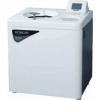
 Hitachi日立CT15RE台式离心机型号: CT15RE
 -
 Hitachi日立超速离心机CP70ME型号: CP70ME
 -
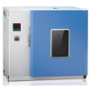 电热鼓风干燥箱型号: CS101-ABN
 -
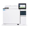 Agilent GC 8890 气相色谱系统型号: 8890 GC
 -
 上海辰华电化学工作站CHI760E型号: CHI760E
 -
 Quantum Design-FusionScope扫描电镜——SEM+AFM二合一显微镜型号: FusionScope
 -
 ASML 光刻机 1460K型号: ASML 光刻机 1460K
 -
 珀金埃尔默PerkinElmer NexION 1100/1000G ICP-MS型号: NexION 1100/1000G ICP-MS


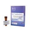
Anti-Rad21 antibody
种属反应性Human,Mouse,Rat
验证应用WB,ICC,IHC-P
抗体类型兔多抗
免疫原Recombinant protein within human Rad21 aa 250-440.
偶联Non-conjugated
Anti-Rad21 antibody性能
形态Liquid
浓度1 mg/ml.
存放说明Store at +4℃ after thawing. Aliquot store at -20℃. Avoid repeated freeze / thaw cycles.
存储缓冲液1*PBS (pH7.4), 0.2% BSA, 50% Glycerol. Preservative: 0.05% Sodium Azide.
亚型IgG
纯化方式Protein affinity purified.
亚细胞定位Centromere, Chromosome, Nucleus.
其它名称
moreAnti-Rad21 antibody应用
WB:1:500-1:2,000
ICC:1:50-1:200
IHC-P:1:100-1:400
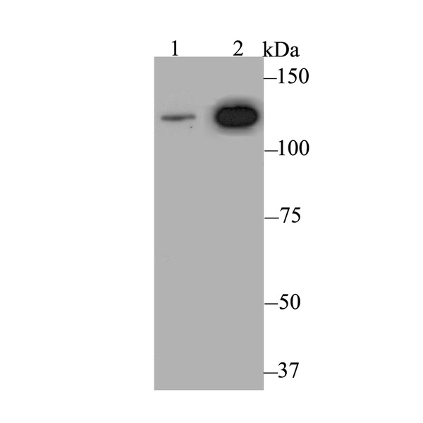
Fig1: Western blot analysis of Rad21 on different lysates. Proteins were transferred to a PVDF membrane and blocked with 5% BSA in PBS for 1 hour at room temperature. The primary antibody was used at a 1:500 dilution in 5% BSA at room temperature for 2 hours. Goat Anti-Rabbit IgG - HRP Secondary Antibody (HA1001) at 1:5,000 dilution was used for 1 hour at room temperature.
Positive control:
Lane 1: Mouse ovary tissue lysate
Lane 2: Daudi cell lysate
Fig2: ICC staining Rad21 in MG-63 cells (green). Formalin fixed cells were permeabilized with 0.1% Triton X-100 in TBS for 10 minutes at room temperature and blocked with 1% Blocker BSA for 15 minutes at room temperature. Cells were probed with Rad21 polyclonal antibody at a dilution of 1:100 for 1 hour at room temperature, washed with PBS. Alexa Fluorc™ 488 Goat anti-Rabbit IgG was used as the secondary antibody at 1/100 dilution. The nuclear counter stain is DAPI (blue).

Fig3: ICC staining Rad21 in SiHa cells (green). Formalin fixed cells were permeabilized with 0.1% Triton X-100 in TBS for 10 minutes at room temperature and blocked with 1% Blocker BSA for 15 minutes at room temperature. Cells were probed with Rad21 polyclonal antibody at a dilution of 1:100 for 1 hour at room temperature, washed with PBS. Alexa Fluorc™ 488 Goat anti-Rabbit IgG was used as the secondary antibody at 1/100 dilution. The nuclear counter stain is DAPI (blue).

Fig4: ICC staining Rad21 in SK-Br-3 cells (green). Formalin fixed cells were permeabilized with 0.1% Triton X-100 in TBS for 10 minutes at room temperature and blocked with 1% Blocker BSA for 15 minutes at room temperature. Cells were probed with Rad21 polyclonal antibody at a dilution of 1:100 for 1 hour at room temperature, washed with PBS. Alexa Fluorc™ 488 Goat anti-Rabbit IgG was used as the secondary antibody at 1/100 dilution. The nuclear counter stain is DAPI (blue).

Fig5: Immunohistochemical analysis of paraffin-embedded rat brain tissue using anti-Rad21 antibody. The section was pre-treated using heat mediated antigen retrieval with sodium citrate buffer (pH 6.0) for 20 minutes. The tissues were blocked in 5% BSA for 30 minutes at room temperature, washed with ddH2O and PBS, and then probed with the antibody (ER1803-73) at 1/200 dilution, for 30 minutes at room temperature and detected using an HRP conjugated compact polymer system. DAB was used as the chrogen. Counter stained with hematoxylin and mounted with DPX.

Fig6: Immunohistochemical analysis of paraffin-embedded human thyroid gland cancer tissue using anti-Rad21 antibody. The section was pre-treated using heat mediated antigen retrieval with sodium citrate buffer (pH 6.0) for 20 minutes. The tissues were blocked in 5% BSA for 30 minutes at room temperature, washed with ddH2O and PBS, and then probed with the antibody (ER1803-73) at 1/200 dilution, for 30 minutes at room temperature and detected using an HRP conjugated compact polymer system. DAB was used as the chrogen. Counter stained with hematoxylin and mounted with DPX.

Fig7: Immunohistochemical analysis of paraffin-embedded human colon tissue using anti-Rad21 antibody. The section was pre-treated using heat mediated antigen retrieval with sodium citrate buffer (pH 6.0) for 20 minutes. The tissues were blocked in 5% BSA

Fig8: Immunohistochemical analysis of paraffin-embedded mouse testis tissue using anti-Rad21 antibody. The section was pre-treated using heat mediated antigen retrieval with sodium citrate buffer (pH 6.0) for 20 minutes. The tissues were blocked in 5% BSA
特别提示:本公司的所有产品仅可用于科研实验,严禁用于临床医疗及其他非科研用途!