


Anti-NCAM-L1 antibody
种属反应性Human,Mouse,Rat
验证应用WB,ICC,IHC-P,FC
抗体类型兔多抗
免疫原Synthetic peptide (KLH-coupled) corresponding to a region of C-terminal residues of NCAM-L1.
偶联Non-conjugated
Anti-NCAM-L1 antibody性能
形态Liquid
浓度1 mg/mL.
存放说明Store at +4℃ after thawing. Aliquot store at -20℃. Avoid repeated freeze / thaw cycles.
存储缓冲液1*PBS (pH7.4), 0.2% BSA, 40% Glycerol. Preservative: 0.05% Sodium Azide.
亚型IgG
纯化方式Peptide affinity purified
亚细胞定位Cell membrane. Cell projection. Membrane.
数据链接P32004 Human
P11627 Mouse其它名称
moreAnti-NCAM-L1 antibody应用
WB: 1:500-1:1,000
ICC: 1:50-1:200
IHC-P: 1:50-1:200
FC: 1:50-1:100
Fig1: Western blot analysis of NCAML1 on human fetal brain tissue lysate using anti-NCAML1 antibody at 1/1,000 dilution.

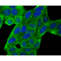
Fig2: ICC staining NCAML1 in 293T cells (green). The nuclear counter stain is DAPI (blue). Cells were fixed in paraformaldehyde, permeabilised with 0.25% Triton X100/PBS.

Fig3: ICC staining NCAML1 in Hela cells (green). The nuclear counter stain is DAPI (blue). Cells were fixed in paraformaldehyde, permeabilised with 0.25% Triton X100/PBS.

Fig4: Immunohistochemical analysis of paraffin-embedded human breast cancer tissue using anti-NCAML1 antibody. Counter stained with hematoxylin.

Fig5: Immunohistochemical analysis of paraffin-embedded human kidney tissue using anti-NCAML1 antibody. Counter stained with hematoxylin.

Fig6: Immunohistochemical analysis of paraffin-embedded mouse kidney tissue using anti-NCAML1 antibody. Counter stained with hematoxylin.

Fig7: Flow cytometric analysis of SHG-44 cells with NCAML1 antibody at 1/100 dilution (red) compared with an unlabelled control (cells without incubation with primary antibody; black). Alexa Fluor 488-conjugated goat anti-rabbit IgG was used as the second
特别提示:本公司的所有产品仅可用于科研实验,严禁用于临床医疗及其他非科研用途!
Anti-NCAM-L1 antibody多少钱?
泽叶生物抗体/抗原的应用资料有吗?
泽叶生物抗体/抗原货号ZY-0805R报价含票含运吗?
泽叶生物货号ZY-0805R货期是多久?
泽叶生物Anti-NCAM-L1 antibody可以申请试用吗?
泽叶生物泽叶生物抗体/抗原售后政策是什么?
Anti-NCAM-L1 antibody有现货吗?