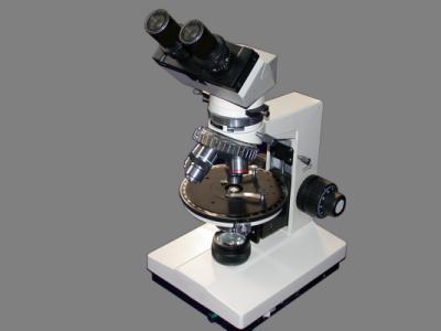
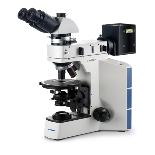
仪器简介:
The polarizing microscope is one of the most useful professional experiment instruments for use in the fields of geology, mineral, and
metallurgical department as well as associated institutions of higher -learning.
This Model AXP-202 Polarizing Microscope can be used for single polarizing examination, orthogonal polarizing examination, and conical polarizing examination as well as for photomicrography.
This microscope is equipped with various accessories such as a gypsum test plate(wavelength), a mica test plate(quarter wavelength),
A quartz wedge and a detachable mechanical stage, etc. having improved performances and fine quality.
技术参数:
2.0 SPECIFICATIONS
2.1 Mechanical tube length:160mm
2.2 objectives: stressless achromatic objectives.
Magnifying power Numerical aperture(N.A) Thickness ofCover glass
4× 0.10
10× 0.25
25× 0.40 0.17
40× 0.65 0.17
63× 0.85 0.17
40× 0.65 0
63× 0.85 0
2.3pieces
Type Magnifying power Diameter ofField of view
Eyepiece with grid 10× 18
Eyepiece with crosshairs 10× 18
Eyepiece with graticule 10× 18
2.4 Final magnifying powers of objectives and eyepieces combination.
Final magnifying powers:
Eyepiece Objectives
4× 10× 25× 40× 63×
10× 40× 100× 250× 400× 630×
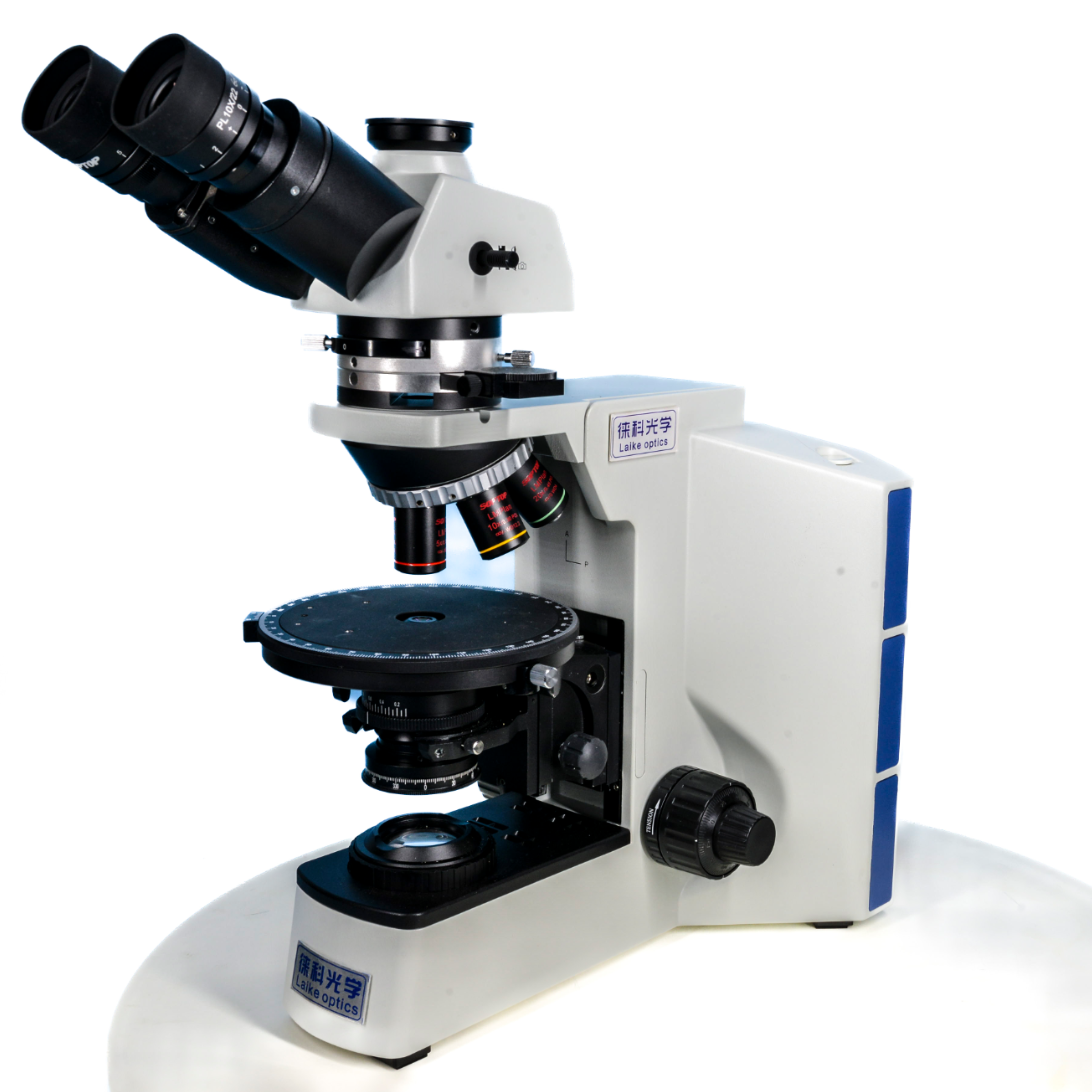
2.5 Quadruple centralized revolving nosepiece for objectives.
2.6 Rotating stage: 140mm in outside dia, calibrated in 360
Degrees, vernier scale 0.1 degree.
2.7 Focussing mechanism
Coaxial, co-guide way coarse and fine focusing adjustment.
Focussing range: 26mm, division 0.002mm.
2.8 Illumination
6V 20W light source, brightness adjustable, both transmitted illumination and vertical illumination are available.
2.9 Line power; 220V/50-60Hz
2.10 Mono-tube viewing head
2.11 35mm photomicrographic equipment, photo-eyepieces:
4×
2.12 Color compensator: one wavelength gypsum red test
plate, a quarter wavelength mica test plate, a quartz wedge.
2.13 Mechanical stage: ranges of movement:30mm×40mm
2.14 Condenser: Abbe two-lens condenser, N.A.=1.25 with
iris diaphragm.
3.0 UNPACKING
Identify the version of the microscope purchased, unpack and bring the microscope out of the case. Cheek up the accessories in the case according to the packing list.
4.0 OPERATION
Insert the plug of the power of this microscope into a suitable grounded 220V AC line power socket. Turn on the power by the switch and the indication light will be lit. Then, switch the changcover switch to the position marked with T(Transmitted) or marked with R(Reflected).
4.1 Place a specimen onto the stage. Make an observation, pay attention to pull the level for pulling the analyzer out from the light path. Open the field iris diaphragm. Make sure that whether field iris diaphragm coincides with the field of view.
Then, close down the iris diaphragm, which should seem to be a circle, with the center of ocular’s cross hairs as its center.
Otherwise, adjust the adjustment screws for the field iris diaphragm to reach the goal. (It is not necessary to make adjustment when using transmitted light.)
4.2 insert the Bertrand lens into the light path, (when reflected illumination is used, allow the magnification marked in the Bertrand lens plate to match with that of objective used), or remove the eyepiece. Look at the bright circular spot at the back focal plane, and open up or close down the aperture iris diaphragm slowly to examine the coincidence of the iris diaphragm and the bright circular spot. Use the method mentioned above in section 4.1 to adjust and eliminate the deviation, if any.
4.3 Place a detachable mechanical stage onto the rotatable stage.
4.4 Secure the specimen properly with the clips of the detachable mechanical stage.
4.5 Adjust the center of the stage and the objective used. Focus the specimen sharply. Find a marked feature in the field of view, and make it to situate at the center of the ocular’s cross hairs. Rotate the stage, if the optical axis of the objective not coincides with the center of the rotation of the stage, then, the target selected will rotate about certain center S(that is the rotating center of the stage), the trajectory of which is a circule. Turn the target point to the point 01, and adjust the center of the objective and allow the point01 to move toward the point S and coincide with it. Then, turn the stage again, to view whether the two points coincide with each other, if any deviation exists, repeat this procedure again.
4.6 When observing the microstructure of a specimen, usually use a low power objective to look for the object, the, move the object towards the center of the field of view, and exchange a high power objective for observation. Care should be taken to avoid the objective being knocked against the specimen and damaged.
When focusing, lift the stage and allow it to close the objective lens as near as possible.
Lower the object surface while viewing. When an image is observed, use the fine adjustment knob for adjustment until a sharp image is obtained.
4.7 Examining the specimen in orthogonal polarized light. As mentioned in section 4.6, when a sharp image is obtained. because the polarizer is still inserted in the optical path., actually it is in a single polarized light. Then insert an analyzer. Pay attention to the graduated marks of the polarizer and analyzer. When both marks are situated at “0” position, it means that that they are in orthogonal. Now, the polarized orientation of the analyzer is in north-south(or up and down).
4.8 Examining the specimen in the conical polarized light. Usually, conical polarized light is used for high power examination. Insert Bertrand lens into the light path when using orthogonal polarizing light. The swinging upper lenses of the condenser are arranged so that they may be moved into the optical axis of the microscope for examining the conical characteristic of the specimen.
4.9 When an indistinct image of the filament of the bulb reveal in the field of view, add a ground glass properly. When the light is yellowish, a filter can be added. (Place a filter above the collector in the base of the microscope in transmitted light).